Explore
 Validate
 Learn
Learn Western blot
Antibody data
- Antibody Data
- Antigen structure
- References [4]
- Comments [0]
- Validations
- Western blot [1]
- Immunocytochemistry [1]
- Immunohistochemistry [1]
- Flow cytometry [1]
Submit
Validation data
Reference
Comment
Report error
- Product number
- MAB513 - Provider product page

- Provider
- R&D Systems
- Product name
- Human MMP-3 Antibody
- Antibody type
- Monoclonal
- Description
- Protein A or G purified from hybridoma culture supernatant. Detects both the pro and active forms of human MMP-3 in direct ELISAs and Western blots. In direct ELISAs, 50-100% cross-reactivity with recombinant mouse MMP-3 is observed, 10% cross-reactivity with recombinant human (rh) MMP-10 is observed and no cross-reactivity with rhMMP-1, -2, -7, -8, -9, -12 or -13 is observed.
- Reactivity
- Human
- Host
- Mouse
- Conjugate
- Unconjugated
- Antigen sequence
P08254- Isotype
- IgG
- Antibody clone number
- 50647
- Vial size
- 500 ug
- Storage
- Use a manual defrost freezer and avoid repeated freeze-thaw cycles. 12 months from date of receipt, -20 to -70 °C as supplied. 1 month, 2 to 8 °C under sterile conditions after reconstitution. 6 months, -20 to -70 °C under sterile conditions after reconstitution.
Submitted references Tissue inhibitors of matrix metalloproteinases in platelets and megakaryocytes: a novel organization for these secreted proteins.
IL-21 is highly produced in Helicobacter pylori-infected gastric mucosa and promotes gelatinases synthesis.
IL-21 is highly produced in Helicobacter pylori-infected gastric mucosa and promotes gelatinases synthesis.
Control of matrix metalloproteinase production in human intestinal fibroblasts by interleukin 21.
Villeneuve J, Block A, Le Bousse-Kerdilès MC, Lepreux S, Nurden P, Ripoche J, Nurden AT
Experimental hematology 2009 Jul;37(7):849-56
Experimental hematology 2009 Jul;37(7):849-56
IL-21 is highly produced in Helicobacter pylori-infected gastric mucosa and promotes gelatinases synthesis.
Caruso R, Fina D, Peluso I, Fantini MC, Tosti C, Del Vecchio Blanco G, Paoluzi OA, Caprioli F, Andrei F, Stolfi C, Romano M, Ricci V, MacDonald TT, Pallone F, Monteleone G
Journal of immunology (Baltimore, Md. : 1950) 2007 May 1;178(9):5957-65
Journal of immunology (Baltimore, Md. : 1950) 2007 May 1;178(9):5957-65
Control of matrix metalloproteinase production in human intestinal fibroblasts by interleukin 21.
Monteleone G, Caruso R, Fina D, Peluso I, Gioia V, Stolfi C, Fantini MC, Caprioli F, Tersigni R, Alessandroni L, MacDonald TT, Pallone F
Gut 2006 Dec;55(12):1774-80
Gut 2006 Dec;55(12):1774-80
No comments: Submit comment
Supportive validation
- Submitted by
- R&D Systems (provider)
- Main image
- Experimental details
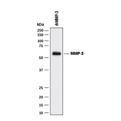
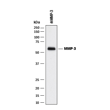
- Detection of Human MMP-3 by Western Blot. Western blot shows Recombinant Human MMP-3 Westrern Blot Standard Protein (2 μL, Catalog # WBC015). PVDF membrane was probed with 1 µg/mL of Mouse Anti-Human MMP-3 Monoclonal Antibody (Catalog # MAB513) followed by HRP-conjugated Anti-Mouse IgG Secondary Antibody (Catalog # HAF018). A specific band was detected for MMP-3 at approximately 55 kDa (as indicated). This experiment was conducted under reducing conditions and using Immunoblot Buffer Group 1.
Supportive validation
- Submitted by
- R&D Systems (provider)
- Main image

- Experimental details
- MMP-3 in MG-63 Human Cell Line. MMP-3 was detected in immersion fixed MG-63 human osteosarcoma cell line using 10 µg/mL Mouse Anti-Human MMP-3 Monoclonal Antibody (Catalog # MAB513) for 3 hours at room temperature. Cells were stained with the NorthernLights™ 557-conjugated Anti-Mouse IgG Secondary Antibody (red; Catalog # NL007) and counterstained with DAPI (blue). View our protocol for Fluorescent ICC Staining of Cells on Coverslips.

Supportive validation
- Submitted by
- R&D Systems (provider)
- Main image

- Experimental details
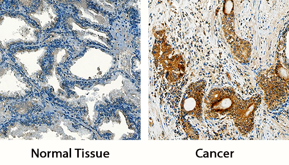
- MMP-3 in Human Prostate and Human Prostate Cancer Tissue. MMP-3 was detected in immersion fixed paraffin-embedded sections of normal human prostate and human prostate cancer tissue using Mouse Anti-Human MMP-3 Monoclonal Antibody (Catalog # MAB513) at 15 µg/mL overnight at 4 °C. Tissue was stained using the Anti-Goat HRP-DAB Cell & Tissue Staining Kit (brown; Catalog # CTS008) and counterstained with hematoxylin (blue). Specific staining was localized to cytoplasm in cancer cells (right panel). View our protocol for Chromogenic IHC Staining of Paraffin-embedded Tissue Sections.
Supportive validation
- Submitted by
- R&D Systems (provider)
- Main image

- Experimental details
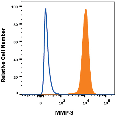
- Detection of MMP-3 in MG-63 Human Cell Line by Flow Cytometry. MG-63 human osteosarcoma cell line was stained with Mouse Anti-Human MMP-3 Monoclonal Antibody (Catalog # MAB513, filled histogram) or isotype control antibody (Catalog # MAB002, open histogram), followed by Allophycocyanin-conjugated Anti-Mouse IgG Secondary Antibody (Catalog # F0101B). To facilitate intracellular staining, cells were fixed with Flow Cytometry Fixation Buffer (Catalog # FC004) and permeabilized with Flow Cytometry Permeabilization/Wash Buffer I (Catalog # FC005). View our protocol for Staining Intracellular Molecules.