Explore
 Validate
 Learn
Learn Western blot
Antibody data
- Antibody Data
- Antigen structure
- References [3]
- Comments [0]
- Validations
- Western blot [1]
- Flow cytometry [1]
Submit
Validation data
Reference
Comment
Report error
- Product number
- AF6775 - Provider product page

- Provider
- R&D Systems
- Product name
- Mouse FCRN Antibody
- Antibody type
- Polyclonal
- Description
- Antigen Affinity-purified. Detects mouse FCRN in direct ELISAs and Western blots. In direct ELISAs, approximately 6% cross-reactivity with recombinant human FCRN is observed.
- Reactivity
- Mouse
- Host
- Goat
- Conjugate
- Unconjugated
- Antigen sequence
AAH03786- Isotype
- IgG
- Vial size
- 100 ug
- Concentration
- LYOPH
- Storage
- Use a manual defrost freezer and avoid repeated freeze-thaw cycles. 12 months from date of receipt, -20 to -70 °C as supplied. 1 month, 2 to 8 °C under sterile conditions after reconstitution. 6 months, -20 to -70 °C under sterile conditions after reconstitution.
Submitted references The Neonatal Fc Receptor and Complement Fixation Facilitate Prophylactic Vaccine-Mediated Humoral Protection against Viral Infection in the Ocular Mucosa.
Distribution of FcRn Across Species and Tissues.
Deletion of Wiskott-Aldrich syndrome protein triggers Rac2 activity and increased cross-presentation by dendritic cells.
Royer DJ, Carr MM, Gurung HR, Halford WP, Carr DJJ
Journal of immunology (Baltimore, Md. : 1950) 2017 Sep 1;199(5):1898-1911
Journal of immunology (Baltimore, Md. : 1950) 2017 Sep 1;199(5):1898-1911
Distribution of FcRn Across Species and Tissues.
Latvala S, Jacobsen B, Otteneder MB, Herrmann A, Kronenberg S
The journal of histochemistry and cytochemistry : official journal of the Histochemistry Society 2017 Jun;65(6):321-333
The journal of histochemistry and cytochemistry : official journal of the Histochemistry Society 2017 Jun;65(6):321-333
Deletion of Wiskott-Aldrich syndrome protein triggers Rac2 activity and increased cross-presentation by dendritic cells.
Baptista MA, Keszei M, Oliveira M, Sunahara KK, Andersson J, Dahlberg CI, Worth AJ, Liedén A, Kuo IC, Wallin RP, Snapper SB, Eidsmo L, Scheynius A, Karlsson MC, Bouma G, Burns SO, Forsell MN, Thrasher AJ, Nylén S, Westerberg LS
Nature communications 2016 Jul 18;7:12175
Nature communications 2016 Jul 18;7:12175
No comments: Submit comment
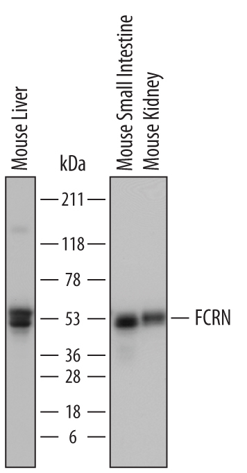
Supportive validation
- Submitted by
- R&D Systems (provider)
- Main image
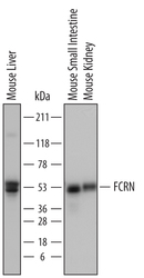
- Experimental details
- Detection of Mouse FCRN by Western Blot. Western blot shows lysates of mouse liver tissue, mouse small intestine tissue, and mouse kidney tissue. PVDF membrane was probed with 0.25 µg/mL of Goat Anti-Mouse FCRN Antigen Affinity-purified Polyclonal Antibody (Catalog # AF6775) followed by HRP-conjugated Anti-Goat IgG Secondary Antibody (Catalog # HAF017). Specific bands were detected for FCRN at approximately 48 to 55 kDa (as indicated). This experiment was conducted under reducing conditions and using Immunoblot Buffer Group 1.
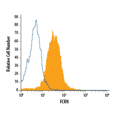
Supportive validation
- Submitted by
- R&D Systems (provider)
- Main image
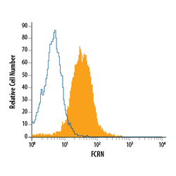
- Experimental details
- Detection of FCRN in J774A.1 Mouse Cell Line by Flow Cytometry. J774A.1 mouse reticulum cell sarcoma macrophage cell line was stained with Goat Anti-Mouse FCRN Antigen Affinity-purified Polyclonal Antibody (Catalog # AF6775, filled histogram) or control antibody (Catalog # AB-108-C, open histogram), followed by Allophycocyanin-conjugated Anti-Goat IgG Secondary Antibody (Catalog # F0108). To facilitate intracellular staining, cells were fixed with paraformaldehyde and permeabilized with saponin.