 Explore
 Validate
 Learn
Learn Immunohistochemistry
Antibody data
- Antibody Data
- Antigen structure
- References [2]
- Comments [0]
- Validations
- Immunohistochemistry [4]
Submit
Validation data
Reference
Comment
Report error
- Product number
- HPA008441 - Provider product page

- Provider
- Atlas Antibodies
- Proper citation
- Atlas Antibodies Cat#HPA008441, RRID:AB_1078983
- Product name
- Anti-GRIA2
- Antibody type
- Polyclonal
- Reactivity
- Human
- Host
- Rabbit
- Conjugate
- Unconjugated
- Antigen sequence
NLRKQRIEISRRGNAGDCLANPAVPWGQGVEIERA
LKQVQVEGLSGNIKFDQNGKRINYTINIMELKTNG
PRKIGYWSEVDKMVVTLTELPSGNDTSGLENKTVV
VTTILESPYVMMKKNHEMLEGNERYEGYCVDLA- Isotype
- IgG
- Vial size
- 100 µl
- Storage
- Store at +4°C for short term storage. Long time storage is recommended at -20°C.
Submitted references Identification of differentially expressed genes according to chemosensitivity in advanced ovarian serous adenocarcinomas: expression of GRIA2 predicts better survival.
Novel markers for enterochromaffin cells and gastrointestinal neuroendocrine carcinomas
Choi CH, Choi JJ, Park YA, Lee YY, Song SY, Sung CO, Song T, Kim MK, Kim TJ, Lee JW, Kim HJ, Bae DS, Kim BG
British journal of cancer 2012 Jun 26;107(1):91-9
British journal of cancer 2012 Jun 26;107(1):91-9
Novel markers for enterochromaffin cells and gastrointestinal neuroendocrine carcinomas
Leja J, Essaghir A, Essand M, Wester K, öberg K, Tötterman T, Lloyd R, Vasmatzis G, Demoulin J, Giandomenico V
Modern Pathology 2008 October;22(2):261-272
Modern Pathology 2008 October;22(2):261-272
No comments: Submit comment
Enhanced validation
Supportive validation
- Submitted by
- Atlas Antibodies (provider)
- Enhanced method
- Orthogonal validation
- Main image

- Experimental details
- Immunohistochemistry analysis in human cerebral cortex and pancreas tissues using Anti-GRIA2 antibody. Corresponding GRIA2 RNA-seq data are presented for the same tissues.
- Sample type
- HUMAN

Supportive validation
- Submitted by
- Atlas Antibodies (provider)
- Main image

- Experimental details
- Immunohistochemical staining of human cerebral cortex shows strong cytoplasmic positivity in neuronal cells.

- Submitted by
- Atlas Antibodies (provider)
- Main image
- Experimental details
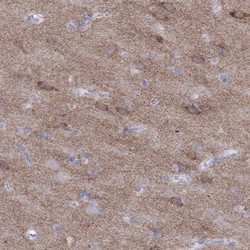
- Immunohistochemical staining of human cerebral cortex shows high expression.
- Sample type
- HUMAN

- Submitted by
- Atlas Antibodies (provider)
- Main image

- Experimental details
- Immunohistochemical staining of human pancreas shows low expression as expected.
- Sample type
- HUMAN
