 Explore
 Validate
 Learn
Learn16488-1-AP
antibody from Invitrogen Antibodies
Targeting: VIL1
D2S1471, VIL
 Western blot Immunocytochemistry
 Immunoprecipitation Immunohistochemistry Flow cytometry Other assay
Antibody data
- Antibody Data
- Antigen structure
- References [0]
- Comments [0]
- Validations
- Western blot [3]
- Immunocytochemistry [1]
- Immunohistochemistry [10]
- Flow cytometry [1]
- Other assay [1]
Submit
Validation data
Reference
Comment
Report error
- Product number
- 16488-1-AP - Provider product page

- Provider
- Invitrogen Antibodies
- Product name
- Villin Polyclonal Antibody
- Antibody type
- Polyclonal
- Antigen
- Other
- Description
- Immunogen sequence: MTKLSAQVK GSLNITTPGL QIWRIEAMQM VPVPSSTFGS FFDGDCYIIL AIHKTASSLS YDIHYWIGQD SSLDEQGAAA IYTTQMDDFL KGRAVQHREV QGNESEAFRG YFKQGLVIRK GGVASGMKHV ETNSYDVQRL LHVKGKRNVV AGEVEMSWKS FNRGDVFLLD LGKLIIQWNG PESTRMERLR GMTLAKEIRD QERGGRTYVG VVDGENELAS PKLMEVMNHV LGKRRELKAA VPDTVVEPAL KAALKLYHVS DSEGNLVVRE VATRPLTQDL LSHEDCYILD QGGLKIYVWK GKKANEQEKK GAMSHALNFI KAKQYPPSTQ VEVQNDGAES AVFQQLFQKW T (1-350 aa encoded by BC017303)
- Reactivity
- Human, Mouse, Rat
- Host
- Rabbit
- Isotype
- IgG
- Vial size
- 150 µL
- Concentration
- 0.13 mg/mL
- Storage
- -20°C
No comments: Submit comment
Supportive validation
- Submitted by
- Invitrogen Antibodies (provider)
- Main image

- Experimental details
- Mouse liver tissue were subjected to SDS PAGE followed by western blot with 16488-1-AP (Villin antibody) at dilution of 1:1000 incubated at room temperature for 1.5 hours.

- Submitted by
- Invitrogen Antibodies (provider)
- Main image

- Experimental details
- Mouse colon tissue were subjected to SDS PAGE followed by western blot with 16488-1-AP (Villin antibody) at dilution of 1:2000 incubated at room temperature for 1.5 hours.

- Submitted by
- Invitrogen Antibodies (provider)
- Main image

- Experimental details
- Mouse kidney tissue were subjected to SDS PAGE followed by western blot with 16488-1-AP (Villin antibody) at dilution of 1:2000 incubated at room temperature for 1.5 hours.

Supportive validation
- Submitted by
- Invitrogen Antibodies (provider)
- Main image

- Experimental details
- Immunofluorescent analysis of COLO 320 cells using 16488-1-AP (Villin antibody) at dilution of 1:50 and Alexa Fluor 488-conjugated AffiniPure Goat Anti-Rabbit IGG (H+L).

Supportive validation
- Submitted by
- Invitrogen Antibodies (provider)
- Main image

- Experimental details
- Immunohistochemistry of paraffin-embedded human small intestine using 16488-1-AP (Villin antibody) at dilution of 1:50 (under 10x lens).

- Submitted by
- Invitrogen Antibodies (provider)
- Main image

- Experimental details
- Immunohistochemistry of paraffin-embedded human small intestine using 16488-1-AP (Villin antibody) at dilution of 1:50 (under 40x lens).

- Submitted by
- Invitrogen Antibodies (provider)
- Main image

- Experimental details
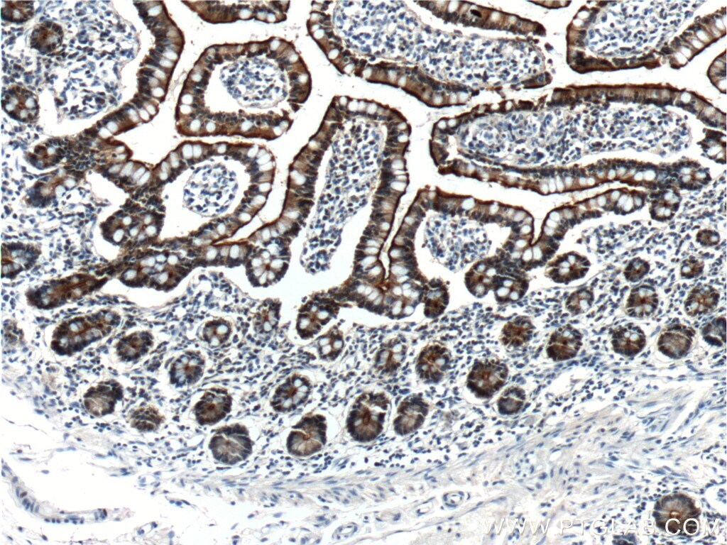
- Immunohistochemistry of paraffin-embedded human colon cancer tissue slide using 16488-1-AP (Villin antibody at dilution of 1:2000 (under 10x lens). heat mediated antigen retrieved with Tris-EDTA buffer (pH 9).

- Submitted by
- Invitrogen Antibodies (provider)
- Main image

- Experimental details
- Immunohistochemistry of paraffin-embedded human colon cancer tissue slide using 16488-1-AP (Villin antibody at dilution of 1:2000 (under 40x lens). heat mediated antigen retrieved with Tris-EDTA buffer (pH 9).

- Submitted by
- Invitrogen Antibodies (provider)
- Main image

- Experimental details
- Immunohistochemistry of paraffin-embedded mouse small intestine tissue slide using 16488-1-AP (Villin antibody at dilution of 1:5000 (under 10x lens). heat mediated antigen retrieved with Tris-EDTA buffer (pH 9).
- Submitted by
- Invitrogen Antibodies (provider)
- Main image

- Experimental details
- Immunohistochemistry of paraffin-embedded mouse small intestine tissue slide using 16488-1-AP (Villin antibody at dilution of 1:5000 (under 40x lens). heat mediated antigen retrieved with Tris-EDTA buffer (pH 9).

- Submitted by
- Invitrogen Antibodies (provider)
- Main image

- Experimental details
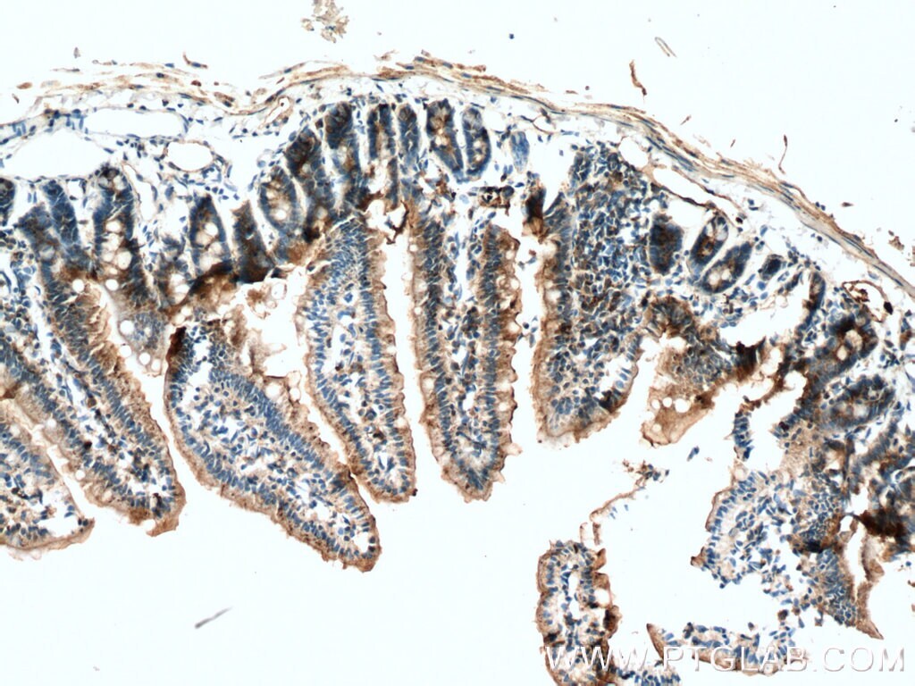
- Immunohistochemistry of paraffin-embedded human small intestine tissue slide using 16488-1-AP (Villin antibody at dilution of 1:5000 (under 10x lens).

- Submitted by
- Invitrogen Antibodies (provider)
- Main image

- Experimental details
- Immunohistochemistry of paraffin-embedded human small intestine tissue slide using 16488-1-AP (Villin antibody at dilution of 1:5000 (under 40x lens).

- Submitted by
- Invitrogen Antibodies (provider)
- Main image

- Experimental details
- Immunohistochemistry of paraffin-embedded human small intestine tissue slide using 16488-1-AP ( Villin antibody at dilution of 1:2000 (under 10x lens).
- Submitted by
- Invitrogen Antibodies (provider)
- Main image

- Experimental details
- Immunohistochemistry of paraffin-embedded human small intestine tissue slide using 16488-1-AP ( Villin antibody at dilution of 1:2000 (under 40x lens).

Supportive validation
- Submitted by
- Invitrogen Antibodies (provider)
- Main image

- Experimental details
- 1X10^6 HepG2 cells were intracellularly stained with 0.2 µg Anti-Human Villin (Product # 16488-1-AP) and CoraLite®488-Conjugated AffiniPure Goat Anti-Rabbit IgG(H+L) at dilution 1:1,000 (red), or 0.2 µg Control Antibody. Cells were fixed with 4% PFA and permeabilized with Flow Cytometry Perm Buffer (PF00011-C).

Supportive validation
- Submitted by
- Invitrogen Antibodies (provider)
- Main image
- Experimental details
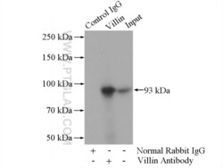
- IP result of anti-Villin (IP:16488-1-AP, 4ug; Detection:16488-1-AP 1:300) with mouse kidney tissue lysate 4000ug.
