 Explore
 Validate
 Learn
Learn Western blot
 Immunocytochemistry
Antibody data
- Antibody Data
- Antigen structure
- References [1]
- Comments [0]
- Validations
- Western blot [9]
- Immunocytochemistry [1]
- Immunoprecipitation [1]
- Immunohistochemistry [3]
Submit
Validation data
Reference
Comment
Report error
- Product number
- GTX109121 - Provider product page

- Provider
- GeneTex
- Proper citation
- GeneTex Cat#GTX109121, RRID:AB_1950421
- Product name
- Glutamine synthetase antibody
- Antibody type
- Polyclonal
- Reactivity
- Human, Mouse, Rat
- Host
- Rabbit
Submitted references FOXO1 activates glutamine synthetase gene in mouse skeletal muscles through a region downstream of 3'-UTR: possible contribution to ammonia detoxification.
Kamei Y, Hattori M, Hatazawa Y, Kasahara T, Kanou M, Kanai S, Yuan X, Suganami T, Lamers WH, Kitamura T, Ogawa Y
American journal of physiology. Endocrinology and metabolism 2014 Sep 15;307(6):E485-93
American journal of physiology. Endocrinology and metabolism 2014 Sep 15;307(6):E485-93
No comments: Submit comment
Enhanced validation
Supportive validation
- Submitted by
- GeneTex (provider)
- Enhanced method
- Genetic validation
- Main image
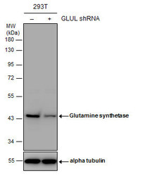
- Experimental details
- Non-transfected (¡V) and transfected (+) 293T whole cell extracts (30 ?g) were separated by 10% SDS-PAGE, and the membrane was blotted with Glutamine synthetase antibody (GTX109121) diluted at 1:1000. The HRP-conjugated anti-rabbit IgG antibody (GTX213110-01) was used to detect the primary antibody.

Supportive validation
- Submitted by
- GeneTex (provider)
- Main image

- Experimental details
- Glutamine synthetase antibody detects GLUL protein by Western blot analysis.A. 30 µg U87-MG whole lysate/extract B. 30 µg SK-N-SH whole cell lysate/extractC. 30 µg IMR32 whole cell lysate/extractD. 30 µg SK-N-AS whole cell lysate/extract 10 % SDS-PAGEGlutamine synthetase antibody (GTX109121) dilution: 1:1000
- Validation comment
- WB

- Submitted by
- GeneTex (provider)
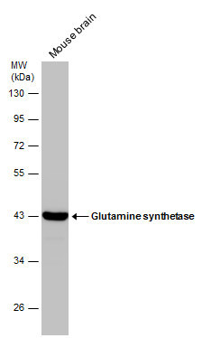
- Main image

- Experimental details
- Sample (20 ug of whole cell lysate) A: mouse brain 10% SDS PAGE GTX109121 diluted at 1:20000
- Validation comment
- WB

- Submitted by
- GeneTex (provider)
- Main image

- Experimental details
- Glutamine synthetase antibody detects GLUL protein by Western blot analysis.A. 50 µg rat brain lysate/extract 10 % SDS-PAGEGlutamine synthetase antibody (GTX109121) dilution: 1:10000
- Validation comment
- WB

- Submitted by
- GeneTex (provider)
- Main image

- Experimental details
- Glutamine synthetase antibody detects Glutamine synthetase protein by western blot analysis. Various whole cell extracts (30 ?g) were separated by 10% SDS-PAGE, and the membrane was blotted with Glutamine synthetase antibody (GTX109121) diluted at a dilution of 1:5000. The HRP-conjugated anti-rabbit IgG antibody (GTX213110-01) was used to detect the primary antibody.

- Submitted by
- GeneTex (provider)
- Main image

- Experimental details
- Mouse tissue extract (50 ?g) was separated by 10% SDS-PAGE, and the membrane was blotted with Glutamine synthetase antibody (GTX109121) diluted at 1:50000. The HRP-conjugated anti-rabbit IgG antibody (GTX213110-01) was used to detect the primary antibody.
- Submitted by
- GeneTex (provider)
- Main image

- Experimental details
- Rat tissue extract (50 ?g) was separated by 10% SDS-PAGE, and the membrane was blotted with Glutamine synthetase antibody (GTX109121) diluted at 1:50000. The HRP-conjugated anti-rabbit IgG antibody (GTX213110-01) was used to detect the primary antibody.

- Submitted by
- GeneTex (provider)
- Main image

- Experimental details
- Mouse tissue extract (50 ?g) was separated by 10% SDS-PAGE, and the membrane was blotted with Glutamine synthetase antibody (GTX109121) diluted at 1:20000. The HRP-conjugated anti-rabbit IgG antibody (GTX213110-01) was used to detect the primary antibody.

- Submitted by
- GeneTex (provider)
- Main image

- Experimental details
- Non-transfected (¡V) and transfected (+) 293T whole cell extracts (30 ?g) were separated by 10% SDS-PAGE, and the membrane was blotted with Glutamine synthetase antibody (GTX109121) diluted at 1:1000. The HRP-conjugated anti-rabbit IgG antibody (GTX213110-01) was used to detect the primary antibody.

Supportive validation
- Submitted by
- GeneTex (provider)
- Main image

- Experimental details
- Glutamine synthetase antibody detects Glutamine synthetase protein at astrocytes by immunofluorescent analysis.Sample: DIV9 rat E18 primary cortical neurons were fixed in 4% paraformaldehyde at RT for 15 min.Green: Glutamine synthetase protein stained by Glutamine synthetase antibody (GTX109121) diluted at 1:500.Red: beta Tubulin 3/ Tuj1, a neuron cell marker, stained by beta Tubulin 3/ Tuj1 antibody [GT11710] (GTX631836) diluted at 1:500.Blue: Fluoroshield with DAPI (GTX30920).

Supportive validation
- Submitted by
- GeneTex (provider)
- Main image

- Experimental details
- Immunoprecipitation of Glutamine synthetase protein from IMR32 whole cell extracts using 5 £gg of Glutamine synthetase antibody (GTX109121).Western blot analysis was performed using Glutamine synthetase antibody (GTX109121).EasyBlot anti-Rabbit IgG (GTX221666-01) was used as a secondary reagent.

Supportive validation
- Submitted by
- GeneTex (provider)
- Main image

- Experimental details
- Immunohistochemical analysis of paraffin-embedded H441 xenograft , using Glutamine Synthetase (GTX109121) antibody at 1:500 dilution.

- Submitted by
- GeneTex (provider)
- Main image

- Experimental details
- Glutamine synthetase antibody detects Glutamine synthetase protein expression by immunohistochemical analysis.Sample: Frozen-sectioned adult mouse cerebellum. Green: Glutamine synthetase protein stained by Glutamine synthetase antibody (GTX109121) diluted at 1:250.Red: beta Tubulin 3/ TUJ1, stained by beta Tubulin 3/ TUJ1 antibody [GT11710] (GTX631836) diluted at 1:500.Blue: Fluoroshield with DAPI (GTX30920).

- Submitted by
- GeneTex (provider)
- Main image

- Experimental details
- Glutamine synthetase antibody detects Glutamine synthetase protein expression by immunohistochemical analysis.Sample:Paraffin-embedded adult mouse retina. Green: Glutamine synthetase protein stained by Glutamine synthetase antibody (GTX109121) diluted at 1:250.Red: beta Tubulin 3/ TUJ1, stained by beta Tubulin 3/ TUJ1 antibody [GT11710] (GTX631836) diluted at 1:250.Blue: Fluoroshield with DAPI (GTX30920).
