 Explore
 Validate
 Learn
Learn Western blot
Antibody data
- Antibody Data
- Antigen structure
- References [0]
- Comments [0]
- Validations
- Western blot [3]
- Immunohistochemistry [1]
- Flow cytometry [1]
Submit
Validation data
Reference
Comment
Report error
- Product number
- ARG55748 - Provider product page

- Provider
- Arigo
- Product name
- anti-p70 S6 Kinase beta antibody
- Antibody type
- Monoclonal
- Antigen
- Purified His-tagged recombinant Human p70 S6 Kinase beta
- Reactivity
- Human, Mouse
- Host
- Mouse
- Isotype
- IgG
- Antibody clone number
- 164CT21.2.2
- Vial size
- 50 µl
- Storage
- For continuous use, store undiluted antibody at 2-8°C for up to a week. For long-term storage, aliquot and store at -20°C or below. Storage in frost free freezers is not recommended. Avoid repeated freeze/thaw cycles. Suggest spin the vial prior to opening.
- Handling
- The antibody solution should be gently mixed before use.
No comments: Submit comment
Supportive validation
- Submitted by
- Arigo (provider)
- Main image

- Experimental details
- Western blot: 35 µg of Jurkat, MCF-7, K562, A549, mouse NIH/3T3 cell line (from left to right) stained with ARG55748 anti-p70 S6 Kinase beta antibody at 1:1000 dilution.

- Submitted by
- Arigo (provider)
- Main image

- Experimental details
- Western blot: 15 µg of Jurkat cell line lysates stained with ARG55748 anti-p70 S6 Kinase beta antibody at 1:1000 dilution.

- Submitted by
- Arigo (provider)
- Main image

- Experimental details
- Western blot: 15 µg of mouse NIH-3T3 cell line lysates stained with ARG55748 anti-p70 S6 Kinase beta antibody at 1:2000 dilution.

Supportive validation
- Submitted by
- Arigo (provider)
- Main image
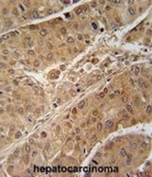
- Experimental details
- Immunohistochemistry: formalin fixed and paraffin embedded human hepatocarcinoma stained with ARG55748 anti-p70 S6 Kinase beta antibody.

Supportive validation
- Submitted by
- Arigo (provider)
- Main image

- Experimental details
- Flow Cytometry: Jurkat cells stained with ARG55748 anti-p70 S6 Kinase beta antibody (right histogram) or without primary antibody control (left histogram), followed by incubation with FITC labelled secondary antibody.
