 Explore
 Validate
 Learn
Learn Western blot
 Other assay
Antibody data
- Antibody Data
- Antigen structure
- References [52]
- Comments [0]
- Validations
- Other assay [26]
Submit
Validation data
Reference
Comment
Report error
- Product number
- 10630D - Provider product page

- Provider
- Invitrogen Antibodies
- Product name
- CD81 Monoclonal Antibody (M38)
- Antibody type
- Monoclonal
- Antigen
- Other
- Description
- The Exosome - anti-CD81 antibody recognizes human CD81, a member of the tetraspanin family, and is predicted to react with feline and rabbit. The antibody has been verified for western blotting of cellular and exosomal CD81 antigen, with a molecular weight of ~25 kDa, and used at 2 µg/mL under non-reducing conditions. Other applications may work but have not been tested.
- Reactivity
- Human
- Host
- Mouse
- Isotype
- IgG
- Antibody clone number
- M38
- Vial size
- 200 µL
- Concentration
- 0.5 mg/mL
- Storage
- 4° C
Submitted references Release of exosomes in polytraumatized patients: The injury pattern is reflected by the surface epitopes.
Analysis of distribution, collection, and confirmation of capacity dependency of small extracellular vesicles toward a therapy for liver cirrhosis.
Searching for airways biomarkers useful to identify progressive pulmonary fibrosis.
Effect of Hypertrophic Scar Fibroblast-Derived Exosomes on Keratinocytes of Normal Human Skin.
In situ hybridization to detect DNA amplification in extracellular vesicles.
Investigating heparin affinity chromatography for extracellular vesicle purification and fractionation.
The Neurotoxicity of Vesicles Secreted by ALS Patient Myotubes Is Specific to Exosome-Like and Not Larger Subtypes.
T-cell trans-synaptic vesicles are distinct and carry greater effector content than constitutive extracellular vesicles.
Effect of Pre-Processing Storage Condition of Cell Culture-Conditioned Medium on Extracellular Vesicles Derived from Human Umbilical Cord-Derived Mesenchymal Stromal Cells.
Inflammatory capacity of exosomes released in the early stages of acute pancreatitis predicts the severity of the disease.
Tracking Radiolabeled Endothelial Microvesicles Predicts Their Therapeutic Efficacy: A Proof-of-Concept Study in Peripheral Ischemia Mouse Model Using SPECT/CT Imaging.
Extracellular vesicles released by non-small cell lung cancer cells drive invasion and permeability in non-tumorigenic lung epithelial cells.
Bone marrow-mesenchymal stem cell-derived extracellular vesicles affect proliferation and apoptosis of leukemia cells in vitro.
The activity of alkaline phosphatase in breast cancer exosomes simplifies the biosensing design.
Usage of celery root exosome as an immune suppressant; Lipidomic characterization of apium graveolens originated exosomes and its suppressive effect on PMA/ionomycin mediated CD4(+) T lymphocyte activation.
Muscle cells of sporadic amyotrophic lateral sclerosis patients secrete neurotoxic vesicles.
Serum Extracellular Vesicle-Derived miR-124-3p as a Diagnostic and Predictive Marker for Early-Stage Acute Ischemic Stroke.
Serum CD203c+ Extracellular Vesicle Serves as a Novel Diagnostic and Prognostic Biomarker for Succinylated Gelatin Induced Perioperative Hypersensitive Reaction.
The membrane associated accessory protein is an adeno-associated viral egress factor.
Single Particle Automated Raman Trapping Analysis of Breast Cancer Cell-Derived Extracellular Vesicles as Cancer Biomarkers.
Mesenchymal growth hormone receptor deficiency leads to failure of alveolar progenitor cell function and severe pulmonary fibrosis.
Downregulation of exosomal let-7d and miR-16 in idiopathic pulmonary fibrosis.
Claudin-2 promotes colorectal cancer liver metastasis and is a biomarker of the replacement type growth pattern.
Plasma cells shape the mesenchymal identity of ovarian cancers through transfer of exosome-derived microRNAs.
Curcumin-primed human BMSC-derived extracellular vesicles reverse IL-1β-induced catabolic responses of OA chondrocytes by upregulating miR-126-3p.
Extracellular Vesicle Surface Signatures in IPF Patients: A Multiplex Bead-Based Flow Cytometry Approach.
Influence of Extracellular Vesicles Isolated From Osteoblasts of Patients With Cox-Arthrosis and/or Osteoporosis on Metabolism and Osteogenic Differentiation of BMSCs.
hBMSC-Derived Extracellular Vesicles Attenuate IL-1β-Induced Catabolic Effects on OA-Chondrocytes by Regulating Pro-inflammatory Signaling Pathways.
Characterization and internalization of small extracellular vesicles released by human primary macrophages derived from circulating monocytes.
Human induced pluripotent stem cells ameliorate hyperoxia-induced lung injury in a mouse model.
Evidence that dysplasia related microRNAs in Barrett's esophagus target PD-L1 expression and contribute to the development of esophageal adenocarcinoma.
Serum small extracellular vesicle-derived LINC00853 as a novel diagnostic marker for early hepatocellular carcinoma.
hnRNPA2B1 inhibits the exosomal export of miR-503 in endothelial cells.
Molecular imaging of extracellular vesicles in vitro via Raman metabolic labelling.
The Potential of CD16 on Plasma-Derived Exosomes as a Liquid Biomarker in Head and Neck Cancer.
Osteosarcoma-Derived Extracellular Vesicles Induce Lung Fibroblast Reprogramming.
Detection of Viral RNA Fragments in Human iPSC-Cardiomyocytes following Treatment with Extracellular Vesicles from SARS-CoV-2 Coding-Sequence-Overexpressing Lung Epithelial Cells.
MicroRNAs in Tumor Exosomes Drive Immune Escape in Melanoma.
Experimental artefacts can lead to misattribution of bioactivity from soluble mesenchymal stem cell paracrine factors to extracellular vesicles.
Gold Nanocluster Extracellular Vesicle Supraparticles: Self-Assembled Nanostructures for Three-Dimensional Uptake Visualization.
Tumor-derived exosomes educate fibroblasts to promote salivary adenoid cystic carcinoma metastasis via NGF-NTRK1 pathway.
Leukemia-derived exosomes and cytokines pave the way for entry into the brain.
Harmonization of exosome isolation from culture supernatants for optimized proteomics analysis.
Helicobacter pylori-induced exosomal MET educates tumour-associated macrophages to promote gastric cancer progression.
Proinflammatory role of blister fluid-derived exosomes in bullous pemphigoid.
Exploring the RNA landscape of endothelial exosomes.
Microvesicle-mediated delivery of miR-1343: impact on markers of fibrosis.
Suppressive effect of an analog of the antimicrobial peptide of LL‑37 on colon cancer cells via exosome‑encapsulated miRNAs.
Exosomal MicroRNA-15a Transfer from the Pancreas Augments Diabetic Complications by Inducing Oxidative Stress.
REG3β modifies cell tumor function by impairing extracellular vesicle uptake.
SCIMP, a transmembrane adaptor protein involved in major histocompatibility complex class II signaling.
Caspase-1 activation of caspase-6 in human apoptotic neurons.
Weber B, Henrich D, Schindler CR, Marzi I, Leppik L
Frontiers in immunology 2023;14:1107150
Frontiers in immunology 2023;14:1107150
Analysis of distribution, collection, and confirmation of capacity dependency of small extracellular vesicles toward a therapy for liver cirrhosis.
Takeda N, Tsuchiya A, Mito M, Natsui K, Natusi Y, Koseki Y, Tomiyoshi K, Yamazaki F, Yoshida Y, Abe H, Sano M, Kido T, Yoshioka Y, Kikuta J, Itoh T, Nishimura K, Ishii M, Ochiya T, Miyajima A, Terai S
Inflammation and regeneration 2023 Oct 9;43(1):48
Inflammation and regeneration 2023 Oct 9;43(1):48
Searching for airways biomarkers useful to identify progressive pulmonary fibrosis.
Soccio P, Moriondo G, Scioscia G, Leo V, Tondo P, Salerno L, Palange P, Barbaro MPF, Lacedonia D
BMC pulmonary medicine 2023 Oct 26;23(1):407
BMC pulmonary medicine 2023 Oct 26;23(1):407
Effect of Hypertrophic Scar Fibroblast-Derived Exosomes on Keratinocytes of Normal Human Skin.
Cui HS, Joo SY, Lee SY, Cho YS, Kim DH, Seo CH
International journal of molecular sciences 2023 Mar 24;24(7)
International journal of molecular sciences 2023 Mar 24;24(7)
In situ hybridization to detect DNA amplification in extracellular vesicles.
Casadei L, Sarchet P, de Faria FCC, Calore F, Nigita G, Tahara S, Cascione L, Wabitsch M, Hornicek FJ, Grignol V, Croce CM, Pollock RE
Journal of extracellular vesicles 2022 Sep;11(9):e12251
Journal of extracellular vesicles 2022 Sep;11(9):e12251
Investigating heparin affinity chromatography for extracellular vesicle purification and fractionation.
Barnes B, Caws T, Thomas S, Shephard AP, Corteling R, Hole P, Bracewell DG
Journal of chromatography. A 2022 May 10;1670:462987
Journal of chromatography. A 2022 May 10;1670:462987
The Neurotoxicity of Vesicles Secreted by ALS Patient Myotubes Is Specific to Exosome-Like and Not Larger Subtypes.
Anakor E, Milla V, Connolly O, Martinat C, Pradat PF, Dumonceaux J, Duddy W, Duguez S
Cells 2022 Mar 1;11(5)
Cells 2022 Mar 1;11(5)
T-cell trans-synaptic vesicles are distinct and carry greater effector content than constitutive extracellular vesicles.
Céspedes PF, Jainarayanan A, Fernández-Messina L, Valvo S, Saliba DG, Kurz E, Kvalvaag A, Chen L, Ganskow C, Colin-York H, Fritzsche M, Peng Y, Dong T, Johnson E, Siller-Farfán JA, Dushek O, Sezgin E, Peacock B, Law A, Aubert D, Engledow S, Attar M, Hester S, Fischer R, Sánchez-Madrid F, Dustin ML
Nature communications 2022 Jun 16;13(1):3460
Nature communications 2022 Jun 16;13(1):3460
Effect of Pre-Processing Storage Condition of Cell Culture-Conditioned Medium on Extracellular Vesicles Derived from Human Umbilical Cord-Derived Mesenchymal Stromal Cells.
Wright A, Snyder OL, Christenson LK, He H, Weiss ML
International journal of molecular sciences 2022 Jul 13;23(14)
International journal of molecular sciences 2022 Jul 13;23(14)
Inflammatory capacity of exosomes released in the early stages of acute pancreatitis predicts the severity of the disease.
Carrascal M, Areny-Balagueró A, de-Madaria E, Cárdenas-Jaén K, García-Rayado G, Rivera R, Martin Mateos RM, Pascual-Moreno I, Gironella M, Abian J, Closa D
The Journal of pathology 2022 Jan;256(1):83-92
The Journal of pathology 2022 Jan;256(1):83-92
Tracking Radiolabeled Endothelial Microvesicles Predicts Their Therapeutic Efficacy: A Proof-of-Concept Study in Peripheral Ischemia Mouse Model Using SPECT/CT Imaging.
Giraud R, Moyon A, Simoncini S, Duchez AC, Nail V, Chareyre C, Bouhlel A, Balasse L, Fernandez S, Vallier L, Hache G, Sabatier F, Dignat-George F, Lacroix R, Guillet B, Garrigue P
Pharmaceutics 2022 Jan 4;14(1)
Pharmaceutics 2022 Jan 4;14(1)
Extracellular vesicles released by non-small cell lung cancer cells drive invasion and permeability in non-tumorigenic lung epithelial cells.
Hasan H, Sohal IS, Soto-Vargas Z, Byappanahalli AM, Humphrey SE, Kubo H, Kitdumrongthum S, Copeland S, Tian F, Chairoungdua A, Kasinski AL
Scientific reports 2022 Jan 19;12(1):972
Scientific reports 2022 Jan 19;12(1):972
Bone marrow-mesenchymal stem cell-derived extracellular vesicles affect proliferation and apoptosis of leukemia cells in vitro.
Phetfong J, Tawonsawatruk T, Kamprom W, Ontong P, Tanyong D, Borwornpinyo S, Supokawej A
FEBS open bio 2022 Feb;12(2):470-479
FEBS open bio 2022 Feb;12(2):470-479
The activity of alkaline phosphatase in breast cancer exosomes simplifies the biosensing design.
Moura SL, Pallarès-Rusiñol A, Sappia L, Martí M, Pividori MI
Biosensors & bioelectronics 2022 Feb 15;198:113826
Biosensors & bioelectronics 2022 Feb 15;198:113826
Usage of celery root exosome as an immune suppressant; Lipidomic characterization of apium graveolens originated exosomes and its suppressive effect on PMA/ionomycin mediated CD4(+) T lymphocyte activation.
Taşlı PN
Journal of food biochemistry 2022 Dec;46(12):e14393
Journal of food biochemistry 2022 Dec;46(12):e14393
Muscle cells of sporadic amyotrophic lateral sclerosis patients secrete neurotoxic vesicles.
Le Gall L, Duddy WJ, Martinat C, Mariot V, Connolly O, Milla V, Anakor E, Ouandaogo ZG, Millecamps S, Lainé J, Vijayakumar UG, Knoblach S, Raoul C, Lucas O, Loeffler JP, Bede P, Behin A, Blasco H, Bruneteau G, Del Mar Amador M, Devos D, Henriques A, Hesters A, Lacomblez L, Laforet P, Langlet T, Leblanc P, Le Forestier N, Maisonobe T, Meininger V, Robelin L, Salachas F, Stojkovic T, Querin G, Dumonceaux J, Butler Browne G, González De Aguilar JL, Duguez S, Pradat PF
Journal of cachexia, sarcopenia and muscle 2022 Apr;13(2):1385-1402
Journal of cachexia, sarcopenia and muscle 2022 Apr;13(2):1385-1402
Serum Extracellular Vesicle-Derived miR-124-3p as a Diagnostic and Predictive Marker for Early-Stage Acute Ischemic Stroke.
Qi Z, Zhao Y, Su Y, Cao B, Yang JJ, Xing Q
Frontiers in molecular biosciences 2021;8:685088
Frontiers in molecular biosciences 2021;8:685088
Serum CD203c+ Extracellular Vesicle Serves as a Novel Diagnostic and Prognostic Biomarker for Succinylated Gelatin Induced Perioperative Hypersensitive Reaction.
Qi Z, Xue Q, Wang H, Cao B, Su Y, Xing Q, Yang JJ
Frontiers in immunology 2021;12:732209
Frontiers in immunology 2021;12:732209
The membrane associated accessory protein is an adeno-associated viral egress factor.
Elmore ZC, Patrick Havlik L, Oh DK, Anderson L, Daaboul G, Devlin GW, Vincent HA, Asokan A
Nature communications 2021 Oct 29;12(1):6239
Nature communications 2021 Oct 29;12(1):6239
Single Particle Automated Raman Trapping Analysis of Breast Cancer Cell-Derived Extracellular Vesicles as Cancer Biomarkers.
Penders J, Nagelkerke A, Cunnane EM, Pedersen SV, Pence IJ, Coombes RC, Stevens MM
ACS nano 2021 Nov 23;15(11):18192-18205
ACS nano 2021 Nov 23;15(11):18192-18205
Mesenchymal growth hormone receptor deficiency leads to failure of alveolar progenitor cell function and severe pulmonary fibrosis.
Xie T, Kulur V, Liu N, Deng N, Wang Y, Rowan SC, Yao C, Huang G, Liu X, Taghavifar F, Liang J, Hogaboam C, Stripp B, Chen P, Jiang D, Noble PW
Science advances 2021 Jun;7(24)
Science advances 2021 Jun;7(24)
Downregulation of exosomal let-7d and miR-16 in idiopathic pulmonary fibrosis.
Lacedonia D, Scioscia G, Soccio P, Conese M, Catucci L, Palladino GP, Simone F, Quarato CMI, Di Gioia S, Rana R, Sollitto F, Foschino-Barbaro MP
BMC pulmonary medicine 2021 Jun 4;21(1):188
BMC pulmonary medicine 2021 Jun 4;21(1):188
Claudin-2 promotes colorectal cancer liver metastasis and is a biomarker of the replacement type growth pattern.
Tabariès S, Annis MG, Lazaris A, Petrillo SK, Huxham J, Abdellatif A, Palmieri V, Chabot J, Johnson RM, Van Laere S, Verhoef C, Hachem Y, Yumeen S, Meti N, Omeroglu A, Altinel G, Gao ZH, Yu ASL, Grünhagen DJ, Vermeulen P, Metrakos P, Siegel PM
Communications biology 2021 Jun 2;4(1):657
Communications biology 2021 Jun 2;4(1):657
Plasma cells shape the mesenchymal identity of ovarian cancers through transfer of exosome-derived microRNAs.
Yang Z, Wang W, Zhao L, Wang X, Gimple RC, Xu L, Wang Y, Rich JN, Zhou S
Science advances 2021 Feb;7(9)
Science advances 2021 Feb;7(9)
Curcumin-primed human BMSC-derived extracellular vesicles reverse IL-1β-induced catabolic responses of OA chondrocytes by upregulating miR-126-3p.
Li S, Stöckl S, Lukas C, Herrmann M, Brochhausen C, König MA, Johnstone B, Grässel S
Stem cell research & therapy 2021 Apr 29;12(1):252
Stem cell research & therapy 2021 Apr 29;12(1):252
Extracellular Vesicle Surface Signatures in IPF Patients: A Multiplex Bead-Based Flow Cytometry Approach.
d'Alessandro M, Soccio P, Bergantini L, Cameli P, Scioscia G, Foschino Barbaro MP, Lacedonia D, Bargagli E
Cells 2021 Apr 28;10(5)
Cells 2021 Apr 28;10(5)
Influence of Extracellular Vesicles Isolated From Osteoblasts of Patients With Cox-Arthrosis and/or Osteoporosis on Metabolism and Osteogenic Differentiation of BMSCs.
Niedermair T, Lukas C, Li S, Stöckl S, Craiovan B, Brochhausen C, Federlin M, Herrmann M, Grässel S
Frontiers in bioengineering and biotechnology 2020;8:615520
Frontiers in bioengineering and biotechnology 2020;8:615520
hBMSC-Derived Extracellular Vesicles Attenuate IL-1β-Induced Catabolic Effects on OA-Chondrocytes by Regulating Pro-inflammatory Signaling Pathways.
Li S, Stöckl S, Lukas C, Götz J, Herrmann M, Federlin M, Grässel S
Frontiers in bioengineering and biotechnology 2020;8:603598
Frontiers in bioengineering and biotechnology 2020;8:603598
Characterization and internalization of small extracellular vesicles released by human primary macrophages derived from circulating monocytes.
Arteaga-Blanco LA, Mojoli A, Monteiro RQ, Sandim V, Menna-Barreto RFS, Pereira-Dutra FS, Bozza PT, Resende RO, Bou-Habib DC
PloS one 2020;15(8):e0237795
PloS one 2020;15(8):e0237795
Human induced pluripotent stem cells ameliorate hyperoxia-induced lung injury in a mouse model.
Mitchell A, Wanczyk H, Jensen T, Finck C
American journal of translational research 2020;12(1):292-307
American journal of translational research 2020;12(1):292-307
Evidence that dysplasia related microRNAs in Barrett's esophagus target PD-L1 expression and contribute to the development of esophageal adenocarcinoma.
Xu J, Yin Z, Yang L, Wu F, Fan J, Huang Q, Jin Y, Yang G
Aging 2020 Sep 9;12(17):17062-17078
Aging 2020 Sep 9;12(17):17062-17078
Serum small extracellular vesicle-derived LINC00853 as a novel diagnostic marker for early hepatocellular carcinoma.
Kim SS, Baek GO, Ahn HR, Sung S, Seo CW, Cho HJ, Nam SW, Cheong JY, Eun JW
Molecular oncology 2020 Oct;14(10):2646-2659
Molecular oncology 2020 Oct;14(10):2646-2659
hnRNPA2B1 inhibits the exosomal export of miR-503 in endothelial cells.
Pérez-Boza J, Boeckx A, Lion M, Dequiedt F, Struman I
Cellular and molecular life sciences : CMLS 2020 Nov;77(21):4413-4428
Cellular and molecular life sciences : CMLS 2020 Nov;77(21):4413-4428
Molecular imaging of extracellular vesicles in vitro via Raman metabolic labelling.
Horgan CC, Nagelkerke A, Whittaker TE, Nele V, Massi L, Kauscher U, Penders J, Bergholt MS, Hood SR, Stevens MM
Journal of materials chemistry. B 2020 May 27;8(20):4447-4459
Journal of materials chemistry. B 2020 May 27;8(20):4447-4459
The Potential of CD16 on Plasma-Derived Exosomes as a Liquid Biomarker in Head and Neck Cancer.
Hofmann L, Ludwig S, Schuler PJ, Hoffmann TK, Brunner C, Theodoraki MN
International journal of molecular sciences 2020 May 26;21(11)
International journal of molecular sciences 2020 May 26;21(11)
Osteosarcoma-Derived Extracellular Vesicles Induce Lung Fibroblast Reprogramming.
Mazumdar A, Urdinez J, Boro A, Migliavacca J, Arlt MJE, Muff R, Fuchs B, Snedeker JG, Gvozdenovic A
International journal of molecular sciences 2020 Jul 30;21(15)
International journal of molecular sciences 2020 Jul 30;21(15)
Detection of Viral RNA Fragments in Human iPSC-Cardiomyocytes following Treatment with Extracellular Vesicles from SARS-CoV-2 Coding-Sequence-Overexpressing Lung Epithelial Cells.
Kwon Y, Nukala SB, Srivastava S, Miyamoto H, Ismail NI, Rehman J, Ong SB, Lee WH, Ong SG
bioRxiv : the preprint server for biology 2020 Jul 1;
bioRxiv : the preprint server for biology 2020 Jul 1;
MicroRNAs in Tumor Exosomes Drive Immune Escape in Melanoma.
Vignard V, Labbé M, Marec N, André-Grégoire G, Jouand N, Fonteneau JF, Labarrière N, Fradin D
Cancer immunology research 2020 Feb;8(2):255-267
Cancer immunology research 2020 Feb;8(2):255-267
Experimental artefacts can lead to misattribution of bioactivity from soluble mesenchymal stem cell paracrine factors to extracellular vesicles.
Whittaker TE, Nagelkerke A, Nele V, Kauscher U, Stevens MM
Journal of extracellular vesicles 2020 Aug 26;9(1):1807674
Journal of extracellular vesicles 2020 Aug 26;9(1):1807674
Gold Nanocluster Extracellular Vesicle Supraparticles: Self-Assembled Nanostructures for Three-Dimensional Uptake Visualization.
Kauscher U, Penders J, Nagelkerke A, Holme MN, Nele V, Massi L, Gopal S, Whittaker TE, Stevens MM
Langmuir : the ACS journal of surfaces and colloids 2020 Apr 14;36(14):3912-3923
Langmuir : the ACS journal of surfaces and colloids 2020 Apr 14;36(14):3912-3923
Tumor-derived exosomes educate fibroblasts to promote salivary adenoid cystic carcinoma metastasis via NGF-NTRK1 pathway.
Xu Z, Zheng X, Zheng J
Oncology letters 2019 Oct;18(4):4082-4091
Oncology letters 2019 Oct;18(4):4082-4091
Leukemia-derived exosomes and cytokines pave the way for entry into the brain.
Kinjyo I, Bragin D, Grattan R, Winter SS, Wilson BS
Journal of leukocyte biology 2019 Apr;105(4):741-753
Journal of leukocyte biology 2019 Apr;105(4):741-753
Harmonization of exosome isolation from culture supernatants for optimized proteomics analysis.
Abramowicz A, Marczak L, Wojakowska A, Zapotoczny S, Whiteside TL, Widlak P, Pietrowska M
PloS one 2018;13(10):e0205496
PloS one 2018;13(10):e0205496
Helicobacter pylori-induced exosomal MET educates tumour-associated macrophages to promote gastric cancer progression.
Che Y, Geng B, Xu Y, Miao X, Chen L, Mu X, Pan J, Zhang C, Zhao T, Wang C, Li X, Wen H, Liu Z, You Q
Journal of cellular and molecular medicine 2018 Nov;22(11):5708-5719
Journal of cellular and molecular medicine 2018 Nov;22(11):5708-5719
Proinflammatory role of blister fluid-derived exosomes in bullous pemphigoid.
Fang H, Shao S, Jiang M, Dang E, Shen S, Zhang J, Qiao P, Li C, Wang G
The Journal of pathology 2018 May;245(1):114-125
The Journal of pathology 2018 May;245(1):114-125
Exploring the RNA landscape of endothelial exosomes.
Pérez-Boza J, Lion M, Struman I
RNA (New York, N.Y.) 2018 Mar;24(3):423-435
RNA (New York, N.Y.) 2018 Mar;24(3):423-435
Microvesicle-mediated delivery of miR-1343: impact on markers of fibrosis.
Stolzenburg LR, Harris A
Cell and tissue research 2018 Feb;371(2):325-338
Cell and tissue research 2018 Feb;371(2):325-338
Suppressive effect of an analog of the antimicrobial peptide of LL‑37 on colon cancer cells via exosome‑encapsulated miRNAs.
Hayashi M, Kuroda K, Ihara K, Iwaya T, Isogai E
International journal of molecular medicine 2018 Dec;42(6):3009-3016
International journal of molecular medicine 2018 Dec;42(6):3009-3016
Exosomal MicroRNA-15a Transfer from the Pancreas Augments Diabetic Complications by Inducing Oxidative Stress.
Kamalden TA, Macgregor-Das AM, Kannan SM, Dunkerly-Eyring B, Khaliddin N, Xu Z, Fusco AP, Yazib SA, Chow RC, Duh EJ, Halushka MK, Steenbergen C, Das S
Antioxidants & redox signaling 2017 Nov 1;27(13):913-930
Antioxidants & redox signaling 2017 Nov 1;27(13):913-930
REG3β modifies cell tumor function by impairing extracellular vesicle uptake.
Bonjoch L, Gironella M, Iovanna JL, Closa D
Scientific reports 2017 Jun 9;7(1):3143
Scientific reports 2017 Jun 9;7(1):3143
SCIMP, a transmembrane adaptor protein involved in major histocompatibility complex class II signaling.
Draber P, Vonkova I, Stepanek O, Hrdinka M, Kucova M, Skopcova T, Otahal P, Angelisova P, Horejsi V, Yeung M, Weiss A, Brdicka T
Molecular and cellular biology 2011 Nov;31(22):4550-62
Molecular and cellular biology 2011 Nov;31(22):4550-62
Caspase-1 activation of caspase-6 in human apoptotic neurons.
Guo H, Pétrin D, Zhang Y, Bergeron C, Goodyer CG, LeBlanc AC
Cell death and differentiation 2006 Feb;13(2):285-92
Cell death and differentiation 2006 Feb;13(2):285-92
No comments: Submit comment
Supportive validation
- Submitted by
- Invitrogen Antibodies (provider)
- Main image

- Experimental details
- NULL

- Submitted by
- Invitrogen Antibodies (provider)
- Main image

- Experimental details
- NULL

- Submitted by
- Invitrogen Antibodies (provider)
- Main image

- Experimental details
- NULL

- Submitted by
- Invitrogen Antibodies (provider)
- Main image

- Experimental details
- NULL

- Submitted by
- Invitrogen Antibodies (provider)
- Main image

- Experimental details
- Figure 1 REG3beta inhibits the uptake of EVs both in vitro and in vivo . ( a ) Transmission electron microscopy images of 120,000 x g pelleted THP1-EVs and MPC-EVs. 2x magnification in the lower right corner to appreciate the double membrane. Scale bars: 200 nm. ( b ) Representative Western blot of EVs samples and cell lysates to confirm the presence of classical exosome markers (CD81, ALIX, TSG101) and the absence of endoplasmic reticulum contamination (Calnexin). ( c,d ) Fluorescence microscopy of THP-1 macrophages ( c ) and MIA PaCa-2 cells (MPC) ( d ) incubated, respectively, with 3 ug/ml of PKH26-labeled MPC EVs or THP1-EVs and increasing concentrations of REG3beta. Nuclei counterstained with DAPI. On the right, quantification of the amount of EVs internalization via fluorimetric reading (n = 4). Data are expressed as mean +- SEM. * P < 0.05, ** P < 0.01, *** P < 0.001, compared to 0 ng/ml REG3beta. ANOVA with Tukey's post-test was used to calculate P -values. Scale bars: 50 um. ( e ) PKH26-labeled EVs injected into tumor xenografts were uptaken by tumor cells (-REG3beta) but remained in the intercellular space when pretreated with REG3beta (+REG3beta). Nuclei counterstained with DAPI. 4x magnification in the top right corner. Scale bars: 50 um.

- Submitted by
- Invitrogen Antibodies (provider)
- Main image

- Experimental details
- Figure 1 Isolation and characterization of exosomes. (A) Image of exosomes observed by transmission electron microscopy. (B) Size distribution of exosomes secreted by non-treated HCT116 cells. (C) Western blot of alpha-tubulin, CD63 and CD81 in exosomes and cells.

- Submitted by
- Invitrogen Antibodies (provider)
- Main image

- Experimental details
- Figure 1 Mesenchymal-epithelial transition factor (MET) expression in AGS cells and cell-derived exosomes. A, Western blotting analysis was used to detect MET protein expression in AGS cells with and without Helicobacter pylori infection for the indicated times. B, qPCR analysis of MET mRNA levels in AGS cells infected with H. pylori for 0, 12 and 24 h. C, The relative expression of Rab27b mRNA was analysed by qPCR in AGS cells infected with H. pylori for 0, 12 and 24 h. * P < 0.05. D, Transmission electron microscopy image of exosomes derived from the AGS cells. Scale bars represent 100 nm. E, Western blotting analysis showing the presence of CD63 and CD81 and the absence of tubulin in AGS cell-derived exosomes. F, MET and p-MET expression in exosomes isolated from AGS cells with and without H. pylori infection was analysed by Western blotting analysis

- Submitted by
- Invitrogen Antibodies (provider)
- Main image

- Experimental details
- Fig 3 Exosome isolation by the size exclusion chromatography. In (a) representative immunoblot showing the distribution of exosome markers (CD63, CD9, CD81) and high-abundance serum proteins (illustrated by Ponceau S staining) in the successive SEC fractions of a FaDU culture medium. In (b) total protein concentrations (mug/uL) in the subsequent SEC fractions. In (c) culture medium supplemented with 5% ED FBS and NOT co-cultured with cells was analyzed as in Panel A; ""E+"" denotes exosome-containing positive control.

- Submitted by
- Invitrogen Antibodies (provider)
- Main image

- Experimental details
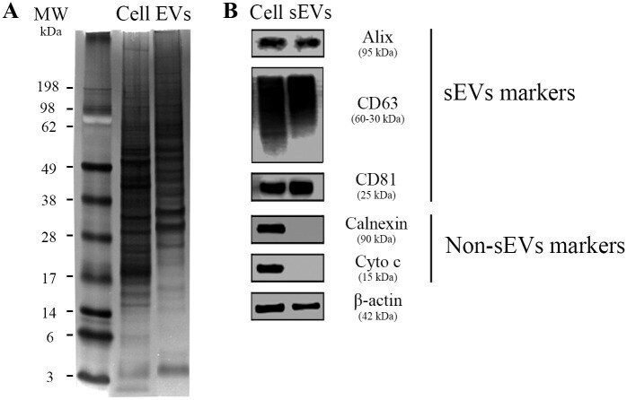
- Fig 4 Protein markers of MDM-derived sEVs. (A) Representative image of three independent assays of polyacrylamide gel stained with silver nitrate after separation of 40 mug total protein from cell (Cell) or sEVs pool lysates (EVs; pools comprise samples from four individual donors). (B) Western blot analysis of sEVs markers (Alix, CD63, and CD81) and non-EVs markers (Calnexin and Cytochrome C) ( n = 3). beta-actin = loading control. 40 mug of total protein were loaded onto the gel. MW: molecular weight marker.
- Submitted by
- Invitrogen Antibodies (provider)
- Main image

- Experimental details
- Figure 1 Characterisation of exosome-like vesicles released from serum by ( A ) transmission electron microscopy, ( B ) dynamic light scattering analysis (133+-25 nm) and ( C ) western blotting. Presence of exosomal markers, CD63, CD9, CD81 and mitochondrial protein cytochrome c in lysates from sera-derived exosomes and cell lysate.

- Submitted by
- Invitrogen Antibodies (provider)
- Main image

- Experimental details
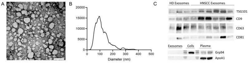
- Figure 1 Characterization of exosomes isolated from plasma. ( A ) Representative transmission electron microscopy (TEM) image of exosomes. Scalebar = 200 nm. ( B ) Representative size distribution of exosomes measured by nanoparticle tracking analysis (NTA). ( C ) Exosomes derived from plasma of healthy donors (HD) and head and neck squamous cell carcinoma (HNSCC) patients were analyzed by Western blot for the presence of exosome specific markers using antibodies against CD63 and CD81 under non-reducing conditions and antibodies against CD9 and TSG101 under reducing conditions. Western blot analysis for the negative marker Grp94 and the apolipoprotein Apo1A was also performed for exosomes, cells, and plasma.
- Submitted by
- Invitrogen Antibodies (provider)
- Main image

- Experimental details
- Figure 1 Characterization of osteosarcoma 143-B-derived extracellular vesicles (EVs) and their uptake by recipient cells. ( A ) 143-B EV size distribution assessed by NanoSight analysis. ( B ) Transmission electron micrographs of 143-B EVs. Scale bar, 250 nm. ( C ) Representative histogram of flow cytometry analysis of exosomal markers CD63 and CD81 in 143-B EV-coated beads. ( D ) Western blot analysis of EV markers (ALIX, CD9) and a cellular marker (calreticulin) in 143-B whole cell protein extracts and in eluted EV fractions from iodixanol based density gradient ultracentrifugation of 143-B EVs. Internalization of PKH67-labelled 143-B EVs by human umbilical vein endothelial cells (HUVEC) ( E ), WI-38 ( F ) and MRC-5 ( G ) cells, as measured by flow cytometry. Representative histograms indicating the percentage of cells internalizing EVs (10 ug) over time (left panels) and the quantification of mean fluorescence intensity (MFI) indicating dose- and time-dependent EV uptake (right panels). The data represent means +- SEM from three independent experiments.

- Submitted by
- Invitrogen Antibodies (provider)
- Main image

- Experimental details
- Fig. 3 Characterization of control EVs and Cur-EVs. a Representative western blot image showing bands of standard surface markers (CD9, CD63, CD81) of control EV- and Cur-EV lysates. b , c Morphology of control EVs and Cur-EVs was monitored by TEM; scale bar, 100 nm. d , e Particle size distribution of control EVs and Cur-EVs was measured by NTA. f , g Quantitative comparison between control EVs and Cur-EVs in count and size measured by NTA; n = 3. h Absorbance at 420 nm of control EVs and Cur-EVs was determined with by spectrophotometry indicating presence of curcumin in Cur-EVs; n = 3. i Cell nuclei were stained with DAPI (blue) and chondrocytes were stained with Phalloidin (green) to visualize the structure of the cytoskeleton. PKH26-labeled control EVs (red) and Cur-EVs (red) were internalized by chondrocytes and visualized with fluorescent microscopy

- Submitted by
- Invitrogen Antibodies (provider)
- Main image

- Experimental details
- Figure 2 Western Blot results. Characterization of isolated extracellular vesicles by Western Blotting analysis using primary antibodies directed to CD81, Alix and loading control beta-actin. Bands were obtained using an exposure of 100 s.

- Submitted by
- Invitrogen Antibodies (provider)
- Main image

- Experimental details
- Fig. 6 Claudin-2 is enriched in extracellular vesicles isolated from patients with replacement-type colorectal cancer liver metastases. a Nanoparticle tracking analysis of EVs detected in plasma derived from either a RHGP liver lesion bearing patient or a DHGP liver lesion bearing patient. b Representative immunoblot analysis of Claudin-2, Claudin-4, CD81, EpCAM, and TSG101 using lysates prepared from EVs isolated from patients with the indicated lesions. c Claudin-2 levels in EVs concentrated from plasma of liver-metastatic CRC patients with the indicated lesions were normalized to TSG101 levels. d Immunoblot analysis of Claudin-2 and TSG101 using lysates prepared from EVs concentrated from patients, pre- and post-resection, with the indicated lesions. e Claudin-2 levels in EVs concentrated from plasma, pre- and post-resection, of liver-metastatic CRC patients with the indicated lesions were normalized to TSG101 levels.

- Submitted by
- Invitrogen Antibodies (provider)
- Main image

- Experimental details
- Fig. 4 ALIX and CD81 Western Blot analysis. Bands were obtained using an exposure time of 100 s

- Submitted by
- Invitrogen Antibodies (provider)
- Main image

- Experimental details
- FIGURE 2 Characterization of control EVs and gene analysis of hsa-124-3p in serum-derived EVs and serum. (A) Representative image of Western blotting showed bands of standard surface markers ( CD9 , CD63 , and CD81 ) of EVs and EV-depleted serum. (B) Morphology of EVs was monitored by TEM; scale bar: 100 nm. (C,D) Particle size distribution of EVs was determined by NTA. (E,F) Gene expression of hsa-miR-124-3p in EVs and serum of different samples at different time points. Compared to the control group: * p < 0.05; ** p < 0.01; *** p < 0.001; **** p < 0.0001, # Difference between groups: # p < 0.05; ## p < 0.01; #### p < 0.0001; one-way ANOVA with the Newman-Keuls multiple comparison test.

- Submitted by
- Invitrogen Antibodies (provider)
- Main image

- Experimental details
- Figure 1 Characterization of control EVs and HR-EVs. EVs were processed for western blot, TEM and NTA after isolated from 500mul serum of control and HR groups. (A) The same amount (30mul) of control EVs, HR-EVs and EVs-depleted-Serum were identified using western blot. Representative western blot image showing bands of standard surface markers (CD9, CD63, CD81) of control EVs and HR-EVs. (B, C) Morphology of control EVs and HR-EVs was monitored by TEM; Scale bar: 100 nm. (D, E) Particle size distribution of control EVs and HR-EVs was determined by NTA. Distribution of EVs with a size of 90-200 nm in diameter in both groups. (F, G) Quantitative comparison between control EVs and HR-EVs in count and size measured by NTA; n = 10; All values represent mean +- standard deviation. * p < 0,05, independent two-tailed Student's t -tests.

- Submitted by
- Invitrogen Antibodies (provider)
- Main image

- Experimental details
- Figure 2 HR promoted the concentration of EVs and CD203c + -EVs and CD63 + -EVs derived from serum. 500mul serum from control and different time point HR groups were used to pellet EVs, finally, the EVs were resuspended in 200mul PBS. The total concentration of EVs was determined by a BCA assay. The protein expression levels of CD63, CD203c, CD9 and CD81 in EVs/serum were identified using western blotting. (A) Total concentration of EVs isolated from same volume serum of control and different time point HR groups, n = 10. (B-F) Relative CD63, CD203c, CD9 and CD81 protein expression levels of EVs derived from same volume of serum in control and HR groups, 30mul EVs/lane, n = 5 (G-I) Relative CD63 and CD203c expression levels in same volume of serum in the control and HR groups, 30mul serum/lane, n = 5. All values represent the mean +- standard deviation. Difference to control: ** p < 0,01; *** p < 0,001; # Difference between groups: # p < 0,05; ## p < 0,01; 1 way ANOVA with Newman-Keuls Multiple Comparison Test.

- Submitted by
- Invitrogen Antibodies (provider)
- Main image

- Experimental details
- FIGURE 5 EV characterization and validation. (A) Representative SEM pictures of BMSC EVs. White arrows label EVs with 97 nm (left image) and 115 nm (right image) diameter. Magnification: 60,000x. (B) Representative TEM pictures of BMSC EVs. Scale bar left image = 200 nm, magnification: 100,000x. Right image shows magnification of the EV in the green box. The size of objects was determined (red lines). Vertical line = 104 nm, horizontal line = 113 nm. (C) Western Blot analysis of EV specific surface markers CD9 (left image) and CD81 (right image) in BMSC, CA, CA/OP, and OP EVs and in the EV-depleted FCS depl - uc . Lane 1 = MW ladder, Lane 2 = 5 mug; Lanes 3-6 = 8.2 mug; Lane 7 = 10 mul; Exposure time: left image = 3 min, right image = 10 min (Pierce femto Kit). For respective Ponceau Red images, see Supplementary Figure 3 . (D) Test for EV uptake of BMSC-derived EVs into cultured BMSCs using PKH-26 (red) stained EVs (lower panel). PBS solution + PKH-26 stain was used as negative control (upper panel). Nuclei were counterstained with DAPI. Scale bar 100 mum.

- Submitted by
- Invitrogen Antibodies (provider)
- Main image

- Experimental details
- Figure 2 Characterization of hBMSC-derived EVs. (A) Representative western blot image ( n = 4) demonstrates standard surface markers (CD9, CD63, and CD81) of hBMSC-derived EVs (lane 1: hBMSC lysate; lane 2: hBMSC-EVs). (B) Particle size distribution of hBMSC-EVs was measured by NTA. (C) Morphology of hBMSC-EVs was monitored by SEM, scale bar: 1 mum. (D) Cell nuclei were stained with DAPI (blue) and chondrocytes were stained with phalloidin (green) to visualize the cytoskeleton. PKH26-labeled hBMSC-derived EVs (red) internalized by chondrocytes were visualized with fluorescent microscopy.

- Submitted by
- Invitrogen Antibodies (provider)
- Main image

- Experimental details
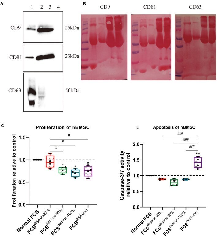
- Figure 3 Characterization of EV-depleted FCS (FCS depl-uc ). Prior to ultracentrifugation, normal FCS was diluted in culture medium to different concentrations (20, 50%, and undiluted = 100%); different FCS depl-uc groups were identified after ultracentrifugation. (A,B) FCS depl-uc-20% was controlled for EV surface makers (CD9, CD63, and CD81) detected by western blotting (lane 1: hBMSC lysate; lane 2: hBMSC-EVs; lane 3: undepleted FCS; lane 4: FCS depl-uc-20% ). Representative western blot image (A) and Ponceau Red-stained images for each surface marker (B) are shown; n = 3. (C,D) Proliferation and apoptosis of hBMSC were determined by BrdU assay and caspase-3/7 activity assay separately after being incubated in culture medium supplemented with the different FCS groups for 24 h. All values represent mean +- standard deviation. *Significant difference to control: * p < 0.05; ** p < 0.01; # Significant difference between groups: # p < 0.05; ### p < 0.001 one-way ANOVA with Newman-Keuls Multiple Comparison Test; n = 4.
- Submitted by
- Invitrogen Antibodies (provider)
- Main image

- Experimental details
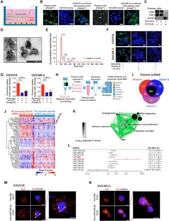
- Fig. 3 Plasma cell exosomal miR-330-3p could be transferred to ovarian cancer. ( A ) Schematic diagram of plasma cells and ovarian cancer cells cocultured in six-well plates. ( B ) Ovarian cancer cells were cocultured in the absence or presence of primary PKH67-labeled plasma cells (green). Nuclei were stained with DAPI (blue) ( n = 3 for each group). Scale bar, 20 mum. ( C ) Western blot analysis for CD63, CD81, and beta-actin in plasma cell exosomes ( n = 3 for each group). ( D ) Electron micrograph of plasma cell exosomes shows the morphological size (50 to 200 nm). Scale bar, 100 nm. ( E ) Size distribution of exosomes was measured using NanoSight analysis. ( F and G ) Immunofluorescent images of PKH67 abrogation in ovarian cancer cells with respective treatment. Scale bar, 20 mum. Statistical chart was plotted ( n = 3 for each group). ( H ) Scheme chart for small RNA sequencing in plasma cell exosomes in patients with ovarian cancer. tRNA, transfer RNA; rRNA, ribosomal RNA; snRNA, small nuclear RNA; snoRNA, small nucleolar RNA; piRNA, Piwi-interacting RNA. ( I ) Venn diagram for overlapped miRNAs identified in ovarian cancer plasma cell exosomes. ( J ) Heatmap for unsupervised hierarchical clustering of GSE73582 dataset using plasma cell exosome-specific miRNA panel as classifiers. ( K ) Cellular programs enriched by GSEA for plasma cell exosome-specific miRNAs represented using Enrichment Map. ( L ) The univariate regression analyses of the identified top miRNAs associa
- Submitted by
- Invitrogen Antibodies (provider)
- Main image

- Experimental details
- Fig. 5 MAAP promotes association of AAV with EVs. A Schematic of EV isolation by iodixanol density gradient from HEK293 suspension culture. B Immunoblots of iodixanol fractions from suspension cells producing recombinant MAAP8Delta vector complemented in trans with CMV-HA and C CMV-MAAP8-HA. EVs, capsid and MAAP were analyzed from the media of HEK293 producing cells at day 3 post transfection. Capsid and MAAP proteins were analyzed by SDS-PAGE under reducing conditions while EV markers (CD81, CD63, CD9) were analyzed by SDS-PAGE under non-reducing conditions ( n = 2). D Graph displaying percent vector genome titer relative to total viral genomes for each fraction from iodixanol gradient purified MAAP8Delta vector complemented in trans with CMV-HA and CMV-MAAP8-HA. E Schematic of EV isolation by size exclusion chromatography (SEC) from HEK293 suspension culture. F Immunoblots of SEC fractions from suspension cells producing recombinant MAAP8Delta vector complemented in trans with CMV-HA and G CMV-MAAP8-HA. EVs and MAAP were analyzed from the media of HEK293 producing cells at day 3 post transfection. MAAP protein was analyzed by SDS-PAGE under reducing conditions while the EV marker CD63 was analyzed by SDS-PAGE under non-reducing conditions ( n = 2). H Graph displaying percent vector genome titer relative to total viral genomes for each fraction from SEC purified MAAP8Delta vector complemented in trans with CMV-HA and CMV-MAAP8-HA.

- Submitted by
- Invitrogen Antibodies (provider)
- Main image

- Experimental details
- MuVs and lmEVs present different markers and have different buoyant properties. ( A ) histograms showing the MuV and lmEV particle-size distributions (representative sample, from ALS EVs). ( B ) Representative Western blots showing the detection of CD63, CD81, CD82, AnnexinA1, ARF6, and actin, in MuVs (line1), lmEVs (line 2) and cells (line 3). Exosomal markers were enriched in MuVs and at relatively low or undetectable levels in lmEVs (EVs were extracted from the same cell culture medium for both exosomal and microparticle markers). Protein loaded on the gel is also shown, as loading control. Cellular contamination was not observed as neither of the vesicle fractions were positive for alpha-skeletal actin. ( C ) Vesicle extracts loaded on iodixanol gradients. MuVs presented classic exosomal buoyant properties while the buoyant range of lmEVs extended to a higher iodixanol density. Top panel: representative Western blot showing detection of CD63 for the MuVs at a density of 1.112 g*mL -1 ; bottom panel: representative Western blot (EVs extracted from the same cell culture medium as top panel) showing detection of Annexin A1 and ARF6 for the lmEVs at a density of 1.086-1.112 g*mL -1 and 1.185-1.230 g*mL -1 . Right panel: iodixanol/sucrose density across the 12 fractions; gradient range from 1.048 to 1.263 g*mL -1 . ( D ) Protein concentration in MuVs samples. Protein quantification was below detection sensitivity for lmEVs (ND: not detected). ( E ) Lipid quantification in MuVs

- Submitted by
- Invitrogen Antibodies (provider)
- Main image

- Experimental details
- Figure 1. Exosomes purified from SACC-83 cells are delivered into HPLF cells. (A) Western blotting analysis showing the presence of CD63, CD81, TSG101 and the absence of GRP94 in SACC-83 cell-derived exosomes. (B) Nanoparticle tracking analysis and (C) transmission electron microscopy images showing exosomes isolated from SACC-83 cells. Magnification, x630; scale bar, 20 um. (D) Confocal microscopy images showing the internalization of PKH67-labeled exosomes in HPLF cells at different incubation times. Scale bar, 5 um. (E) Confocal microscopy images showing the delivery of 0, 25, 50 or 75 ug of exosomes into HPLF cells after 6 h. Scale bar, 5 um. Magnified views are shown on the right. GRP94, glucose-regulated protein 94; HPLF, human periodontal ligament fibroblast; SACC, salivary adenoid cystic carcinoma.
