Explore
 Validate
 Learn
Learn Western blot
 Immunohistochemistry
Antibody data
- Antibody Data
- Antigen structure
- References [5]
- Comments [0]
- Validations
- Western blot [1]
- Immunohistochemistry [5]
Submit
Validation data
Reference
Comment
Report error
- Product number
- HPA005468 - Provider product page

- Provider
- Atlas Antibodies
- Proper citation
- Atlas Antibodies Cat#HPA005468, RRID:AB_1845062
- Product name
- Anti-ASAH1
- Antibody type
- Polyclonal
- Reactivity
- Human
- Host
- Rabbit
- Conjugate
- Unconjugated
- Antigen sequence
ENSTSYEEAKNLLTKTKILAPAYFILGGNQSGEGC
VITRDRKESLDVYELDAKQGRWYVVQTNYDRWKHP
FFLDDRRTPAKMCLNRTSQENISFETMYDVLSTKP
VLNKLTVYTTLIDVTKGQF- Isotype
- IgG
- Vial size
- 100 µl
- Storage
- Store at +4°C for short term storage. Long time storage is recommended at -20°C.
Submitted references Silencing diacylglycerol kinase-theta expression reduces steroid hormone biosynthesis and cholesterol metabolism in human adrenocortical cells
Acid ceramidase (ASAH1) represses steroidogenic factor 1-dependent gene transcription in H295R human adrenocortical cells by binding to the receptor.
Acid ceramidase (ASAH1) is a global regulator of steroidogenic capacity and adrenocortical gene expression.
Genistein stimulates MCF-7 breast cancer cell growth by inducing acid ceramidase (ASAH1) gene expression.
The cAMP-responsive element binding protein (CREB) regulates the expression of acid ceramidase (ASAH1) in H295R human adrenocortical cells.
Cai K, Lucki N, Sewer M
Biochimica et Biophysica Acta (BBA) - Molecular and Cell Biology of Lipids 2014 April;1841(4):552-562
Biochimica et Biophysica Acta (BBA) - Molecular and Cell Biology of Lipids 2014 April;1841(4):552-562
Acid ceramidase (ASAH1) represses steroidogenic factor 1-dependent gene transcription in H295R human adrenocortical cells by binding to the receptor.
Lucki NC, Li D, Bandyopadhyay S, Wang E, Merrill AH, Sewer MB
Molecular and cellular biology 2012 Nov;32(21):4419-31
Molecular and cellular biology 2012 Nov;32(21):4419-31
Acid ceramidase (ASAH1) is a global regulator of steroidogenic capacity and adrenocortical gene expression.
Lucki NC, Bandyopadhyay S, Wang E, Merrill AH, Sewer MB
Molecular endocrinology (Baltimore, Md.) 2012 Feb;26(2):228-43
Molecular endocrinology (Baltimore, Md.) 2012 Feb;26(2):228-43
Genistein stimulates MCF-7 breast cancer cell growth by inducing acid ceramidase (ASAH1) gene expression.
Lucki NC, Sewer MB
The Journal of biological chemistry 2011 Jun 3;286(22):19399-409
The Journal of biological chemistry 2011 Jun 3;286(22):19399-409
The cAMP-responsive element binding protein (CREB) regulates the expression of acid ceramidase (ASAH1) in H295R human adrenocortical cells.
Lucki N, Sewer MB
Biochimica et biophysica acta 2009 Aug;1791(8):706-13
Biochimica et biophysica acta 2009 Aug;1791(8):706-13
No comments: Submit comment
Enhanced validation
- Submitted by
- Atlas Antibodies (provider)
- Enhanced method
- Orthogonal validation
- Main image
- Experimental details
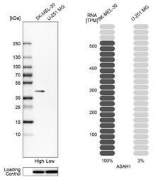
- Western blot analysis in human cell lines SK-MEL-30 and U-251MG using Anti-ASAH1 antibody. Corresponding ASAH1 RNA-seq data are presented for the same cell lines. Loading control: Anti-HDAC1.

Supportive validation
- Submitted by
- Atlas Antibodies (provider)
- Main image
- Experimental details
- Immunohistochemical staining of human epididymis shows strong granular positivity in glandular cells.

- Submitted by
- Atlas Antibodies (provider)
- Main image
- Experimental details
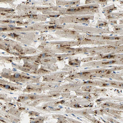
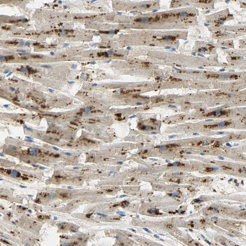
- Immunohistochemical staining of human heart muscle shows strong granular cytoplasmic positivity in cardiomyocytes.
- Sample type
- HUMAN
- Submitted by
- Atlas Antibodies (provider)
- Main image
- Experimental details
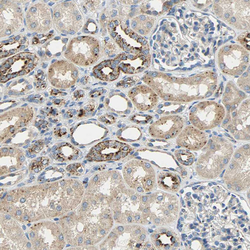
- Immunohistochemical staining of human kidney shows strong granular cytoplasmic positivity in a subset of renal tubules.
- Sample type
- HUMAN

- Submitted by
- Atlas Antibodies (provider)
- Main image
- Experimental details
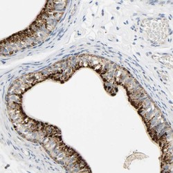
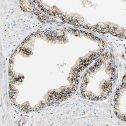
- Immunohistochemical staining of human prostate shows strong granular cytoplasmic positivity in glandular cells.
- Sample type
- HUMAN

- Submitted by
- Atlas Antibodies (provider)
- Main image
- Experimental details
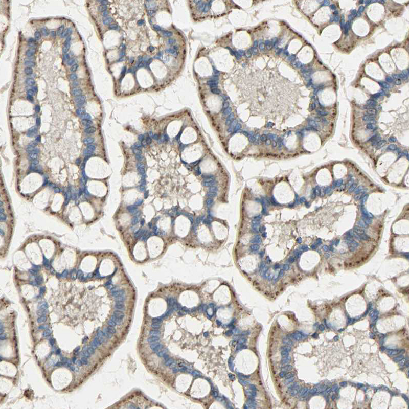
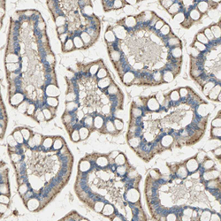
- Immunohistochemical staining of human small intestine shows strong granular cytoplasmic positivity in glandular cells.
- Sample type
- HUMAN