 Explore
 Validate
 Learn
LearnAF1163
antibody from R&D Systems
Targeting: CD40LG
CD154, CD40L, gp39, hCD40L, HIGM1, IMD3, TNFSF5, TRAP
 Western blot
 Immunocytochemistry
Antibody data
- Antibody Data
- Antigen structure
- References [6]
- Comments [0]
- Validations
- Immunocytochemistry [1]
- Blocking/Neutralizing [1]
Submit
Validation data
Reference
Comment
Report error
- Product number
- AF1163 - Provider product page

- Provider
- R&D Systems
- Product name
- Mouse CD40 Ligand/TNFSF5 Antibody
- Antibody type
- Polyclonal
- Description
- Immunogen affinity purified. Detects mouse CD40 Ligand/TNFSF5 in Western blots.
- Reactivity
- Mouse
- Host
- Goat
- Conjugate
- Unconjugated
- Antigen sequence
P27548- Isotype
- IgG
- Vial size
- 100 ug
- Concentration
- LYOPH
- Storage
- Use a manual defrost freezer and avoid repeated freeze-thaw cycles. 12 months from date of receipt, -20 to -70 °C as supplied. 1 month, 2 to 8 °C under sterile conditions after reconstitution. 6 months, -20 to -70 °C under sterile conditions after reconstitution.
Submitted references Identification and characterisation of the CD40-ligand of Sigmodon hispidus.
Vaccination with a fusion protein that introduces HIV-1 gag antigen into a multitrimer CD40L construct results in enhanced CD8+ T cell responses and protection from viral challenge by vaccinia-gag.
The tumor necrosis factor family receptors RANK and CD40 cooperatively establish the thymic medullary microenvironment and self-tolerance.
CD40L expressed from the canarypox vector, ALVAC, can boost immunogenicity of HIV-1 canarypox vaccine in mice and enhance the in vitro expansion of viral specific CD8+ T cell memory responses from HIV-1-infected and HIV-1-uninfected individuals.
Preformed CD40 ligand exists in secretory lysosomes in effector and memory CD4+ T cells and is quickly expressed on the cell surface in an antigen-specific manner.
Reduced pathology following infection with transgenic Leishmania major expressing murine CD40 ligand.
Russell MS, Muralidharan A, Larocque L, Cao J, Deschambault Y, Varga J, Thulasi Raman SN, Li X
PloS one 2018;13(7):e0199067
PloS one 2018;13(7):e0199067
Vaccination with a fusion protein that introduces HIV-1 gag antigen into a multitrimer CD40L construct results in enhanced CD8+ T cell responses and protection from viral challenge by vaccinia-gag.
Gupta S, Termini JM, Raffa FN, Williams CA, Kornbluth RS, Stone GW
Journal of virology 2014 Feb;88(3):1492-501
Journal of virology 2014 Feb;88(3):1492-501
The tumor necrosis factor family receptors RANK and CD40 cooperatively establish the thymic medullary microenvironment and self-tolerance.
Akiyama T, Shimo Y, Yanai H, Qin J, Ohshima D, Maruyama Y, Asaumi Y, Kitazawa J, Takayanagi H, Penninger JM, Matsumoto M, Nitta T, Takahama Y, Inoue J
Immunity 2008 Sep 19;29(3):423-37
Immunity 2008 Sep 19;29(3):423-37
CD40L expressed from the canarypox vector, ALVAC, can boost immunogenicity of HIV-1 canarypox vaccine in mice and enhance the in vitro expansion of viral specific CD8+ T cell memory responses from HIV-1-infected and HIV-1-uninfected individuals.
Liu J, Yu Q, Stone GW, Yue FY, Ngai N, Jones RB, Kornbluth RS, Ostrowski MA
Vaccine 2008 Jul 29;26(32):4062-72
Vaccine 2008 Jul 29;26(32):4062-72
Preformed CD40 ligand exists in secretory lysosomes in effector and memory CD4+ T cells and is quickly expressed on the cell surface in an antigen-specific manner.
Koguchi Y, Thauland TJ, Slifka MK, Parker DC
Blood 2007 Oct 1;110(7):2520-7
Blood 2007 Oct 1;110(7):2520-7
Reduced pathology following infection with transgenic Leishmania major expressing murine CD40 ligand.
Field AE, Wagage S, Conrad SM, Mosser DM
Infection and immunity 2007 Jun;75(6):3140-9
Infection and immunity 2007 Jun;75(6):3140-9
No comments: Submit comment
Supportive validation
- Submitted by
- R&D Systems (provider)
- Main image

- Experimental details
- CD40 Ligand/TNFSF5 in Mouse Splenocytes. CD40 Ligand/TNFSF5 was detected in immersion fixed mouse splenocytes either stimulated with PHA (left panel) or untreated (right panel) using Goat Anti-Mouse CD40 Ligand/TNFSF5 Antigen Affinity-purified Polyclonal Antibody (Catalog # AF1163) at 15 µg/mL for 3 hours at room temperature. Cells were stained using the NorthernLights™ 557-conjugated Anti-Goat IgG Secondary Antibody (red; Catalog # NL001) and counterstained with DAPI (blue). Specific staining was localized to cytoplasm. View our protocol for Fluorescent ICC Staining of Non-adherent Cells.

Supportive validation
- Submitted by
- R&D Systems (provider)
- Main image
- Experimental details
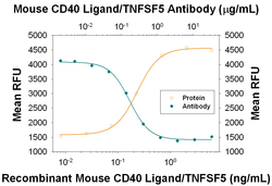
- Proliferation Induced by CD40 Ligand/TNFSF5 and Neutralization by Mouse CD40 Ligand/TNFSF5 Antibody. Recombinant Mouse CD40 Ligand/TNFSF5 (Catalog # 8230-CL) induces proliferation in mouse splenic B cells in a dose-dependent manner (orange line), as measured by Resazurin (Catalog # AR002). Proliferation elicited by Recombinant Mouse CD40 Ligand/TNFSF5 (0.5 ng/mL) is neutralized (green line) by increasing concentrations of Goat Anti-Mouse CD40 Ligand/TNFSF5 Antigen Affinity-purified Polyclonal Antibody (Catalog # AF1163). The ND50 is typically 0.08-0.4 µg/mL.
