 Explore
 Validate
 Learn
Learn Western blot
Antibody data
- Antibody Data
- Antigen structure
- References [1]
- Comments [0]
- Validations
- Western blot [3]
- Immunohistochemistry [1]
- Other assay [1]
Submit
Validation data
Reference
Comment
Report error
- Product number
- PA5-79824 - Provider product page

- Provider
- Invitrogen Antibodies
- Product name
- PKLR Polyclonal Antibody
- Antibody type
- Polyclonal
- Antigen
- Synthetic peptide
- Description
- Reconstitute with 0.2 mL of distilled water to yield a concentration of 500 µg/mL.
- Reactivity
- Human, Mouse, Rat
- Host
- Rabbit
- Isotype
- IgG
- Vial size
- 100 µg
- Concentration
- 500 µg/mL
- Storage
- -20°C
Submitted references Liver Pyruvate Kinase Promotes NAFLD/NASH in Both Mice and Humans in a Sex-Specific Manner.
Chella Krishnan K, Floyd RR, Sabir S, Jayasekera DW, Leon-Mimila PV, Jones AE, Cortez AA, Shravah V, Péterfy M, Stiles L, Canizales-Quinteros S, Divakaruni AS, Huertas-Vazquez A, Lusis AJ
Cellular and molecular gastroenterology and hepatology 2021;11(2):389-406
Cellular and molecular gastroenterology and hepatology 2021;11(2):389-406
No comments: Submit comment
Supportive validation
- Submitted by
- Invitrogen Antibodies (provider)
- Main image

- Experimental details
- Western blot analysis of PKLR in Lane 1: rat liver tissue lysate, Lane 2: mouse liver tissue lysate using 50 µg per well. Sample was incubated with PKLR (Product # PA5-79824) at a dilution of 0.5 µg/mL.

- Submitted by
- Invitrogen Antibodies (provider)
- Main image

- Experimental details
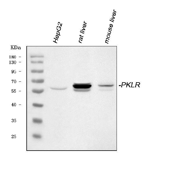
- Western blot analysis of PKLR in, Lane 1: human HepG2 whole cell lysates, Lane 2: rat liver tissue lysates, Lane 3: mouse liver tissue lysates. Electrophoresis was performed on a 5-20% SDS-PAGE gel at 70V (Stacking gel) / 90V (Resolving gel) for 2-3 hours. The sample well of each lane was loaded with 30 µg of sample under reducing conditions. After Electrophoresis, proteins were transferred to a nitrocellulose membrane at 150 mA for 50-90 minutes. The membrane was blocked with 5% non-fat milk/TBS for 1. 5 hour at RT. The membrane was incubated with PKLR Polyclonal Antibody (Product # PA5-79824) at 0.5 μg/mL overnight at 4°C, then washed with TBS-0. 1% Tween 3 times with 5 minutes each and probed with a goat anti-rabbit IgG-HRP secondary antibody at a dilution of 1:5,000 for 1. 5 hour at RT. The signal is developed using an Enhanced Chemiluminescent detection (ECL) kit. A specific band was detected for PKLR at approximately 62 kDa. The expected band size for PKLR is at 62 kDa.
- Submitted by
- Invitrogen Antibodies (provider)
- Main image

- Experimental details
- Western blot was performed using Anti-PKLR Polyclonal Antibody (Product # PA5-79824) and a 58kDa band corresponding to PKLR was observed in Mouse Liver and Rat Liver, but not in the other tissue models which are reported to be negative for PKLR expression. Whole cell lysate (30ug lysate) of Mouse Liver (Lane 1), Mouse Lung (Lane 2), Mouse Brain (Lane 3), Mouse Skeletal muscle (Lane 4), Rat Liver (Lane 5), Rat Lung (Lane 6), Rat Brain (Lane 7), and Rat Skeletal muscle (Lane 8) were electrophoresed using Novex® NuPAGE® 4-12 % Bis-Tris gel (Product # NP0322BOX). Resolved proteins were then transferred onto a nitrocellulose membrane (Product # IB23001) by iBlot® 2 Dry Blotting System (Product # IB21001). The blot was probed with the primary antibody (0.5µg/ml) and detected by chemiluminescence with Goat anti-Rabbit IgG (H+L), Superclonal™ Recombinant Secondary Antibody, HRP conjugate (Product # A27036, 1:4000 dilution) using the iBright FL 1000 (Product # A32752). Chemiluminescent detection was performed using Novex® ECL Chemiluminescent Substrate Reagent Kit (Product # WP20005).

Supportive validation
- Submitted by
- Invitrogen Antibodies (provider)
- Main image

- Experimental details
- Immunohistochemistry analysis of PKLR on paraffin-embedded human intestinal cancer tissue. Sample was incubated with PKLR polyclonal antibody (Product# PA5-79824).

Supportive validation
- Submitted by
- Invitrogen Antibodies (provider)
- Main image

- Experimental details
- Figure 2 AAV-mediated modulation of hepatic L-PK expression. Eight-week-old male C57BL/6J mice were injected with either loss-of-function (shffLuc or shPklr) or gain-of-function (green fluorescent protein or PKLR) AAV vectors under the control of Tbg promoter and fed HF/HS diet for 17 additional weeks. ( A ) qPCR analyses of hepatic Pklr expression and ( B ) immunoblot analyses of hepatic PKLR in L-PK KD and OEx mice. GAPDH was used as a loading control. ( C ) Quantification of average PKLR levels normalized to GAPDH for each group. qPCR analyses of Pklr expression ( D ) 48 hours after siRNA transfection of AML12 cells (experiment was repeated 2 independent times with n = 3 wells per group each time) and ( E ) 12 weeks after NASH diet in L-PK KD mice. Data are presented as mean +- SEM (n = 6-8 livers for RNA and 3 for protein analyses per group). P values were calculated by unpaired Student t test. *** P < .001.
