Explore
 Validate
 Learn
Learn Western blot
Antibody data
- Antibody Data
- Antigen structure
- References [0]
- Comments [0]
- Validations
- Western blot [3]
- Immunocytochemistry [4]
- Immunohistochemistry [2]
- Flow cytometry [1]
Submit
Validation data
Reference
Comment
Report error
- Product number
- MA5-34767 - Provider product page

- Provider
- Invitrogen Antibodies
- Product name
- SENP1 Recombinant Rabbit Monoclonal Antibody (JG37-79)
- Antibody type
- Monoclonal
- Antigen
- Recombinant full-length protein
- Description
- Positive Control: K562, LOVO, SH-SY-5Y, SiHa, Daudi, human liver tissue, human small Intestine tissue.
- Reactivity
- Human, Rat
- Host
- Rabbit
- Isotype
- IgG
- Antibody clone number
- JG37-79
- Vial size
- 100 µL
- Concentration
- 1 mg/mL
- Storage
- -20° C, Avoid Freeze/Thaw Cycles, store in dark
No comments: Submit comment
Supportive validation
- Submitted by
- Invitrogen Antibodies (provider)
- Main image

- Experimental details
- Western blot analysis of SENP1 in K562 cell. Samples were incubated with SENP1 monoclonal antibody (Product # MA5-34767), at a dilution of 1:1000.
- Submitted by
- Invitrogen Antibodies (provider)
- Main image
- Experimental details
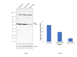
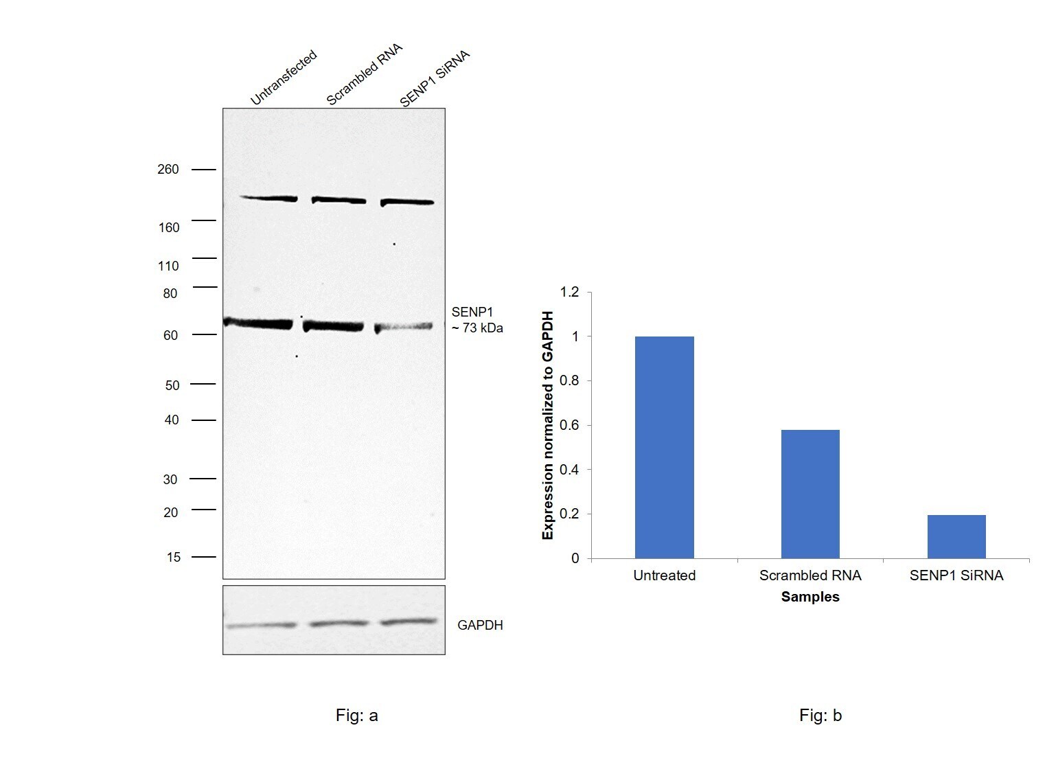
- Knockdown of Senp1; sentrin/SUMO-specific protease SENP1 was achieved by transfecting T-47D with 2Senp1; sentrin/SUMO-specific protease SENP1 specific siRNAs (Silencer® select Product # S26615, S26616). Western blot analysis (Fig. a) was performed using Whole cell extracts from the Senp1; sentrin/SUMO-specific protease SENP1 knockdown cells (lane 3), non-targeting scrambled siRNA transfected cells (lane 2) and untransfected cells (lane 1). The blot was probed with SENP1 Recombinant Rabbit Monoclonal Antibody (JG37-79) (Product # MA5-34767, 1:2000 dilution) and Goat anti-Rabbit IgG (H+L) Superclonal™ Recombinant Secondary Antibody, HRP (Product # A27036, 1:20,000 dilution). Densitometric analysis of this western blot is shown in histogram (Fig. b). Decrease in signal upon siRNA mediated knock down confirms that antibody is specific to Senp1; sentrin/SUMO-specific protease SENP1.
- Submitted by
- Invitrogen Antibodies (provider)
- Main image
- Experimental details
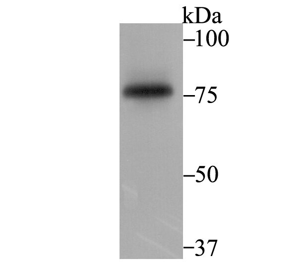
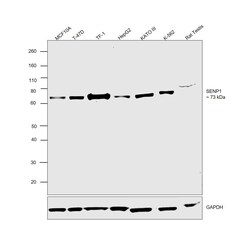
- Western blot was performed using SENP1 Recombinant Rabbit Monoclonal Antibody (JG37-79) (Product # MA5-34767) and a 73 kDa band corresponding to Senp1; sentrin/SUMO-specific protease SENP1 was observed across cell lines and tissue. Whole cell extracts (30 µg lysate) of MCF 10A (Lane 1), T-47D (Lane 2), TF-1 (Lane 3), Hep G2 (Lane 4), KATO III (Lane 5), K-562 (Lane 6) and Rat Testis (Lane 7) were electrophoresed using NuPAGE™ 4-12% Bis-Tris Protein Gel (Product # NP0322BOX), 12 well. Resolved proteins were then transferred onto a nitrocellulose membrane (Product # IB23001) by iBlot® 2 Dry Blotting System (Product # IB21001). The blot was probed with the primary antibody (1:2000 dilution) and detected by chemiluminescence with Goat anti-Rabbit IgG (H+L) Superclonal™ Recombinant Secondary Antibody, HRP (Product # A27036, 1:20,000 dilution) using the iBright™ FL1500 Imaging System (Product # A44115). Chemiluminescent detection was performed using SuperSignal™ West Pico PLUS Chemiluminescent Substrate (Product # 34580).
Supportive validation
- Submitted by
- Invitrogen Antibodies (provider)
- Main image
- Experimental details
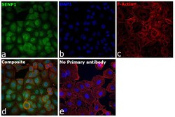
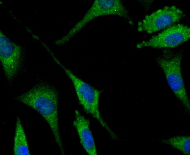
- Immunofluorescence analysis of sentrin/SUMO-specific protease SENP1 was performed using 70% confluent log phase A549 cells. The cells were fixed with 4% paraformaldehyde for 10 minutes, permeabilized with 0.1% Triton™ X-100 for 15 minutes, and blocked with 2% BSA for 1 hour at room temperature. The cells were labeled with SENP1 Recombinant Rabbit Monoclonal Antibody (JG37-79) (Product # MA5-34767) at 1:100 dilution in 0.1% BSA, incubated at 4 degree celsius overnight and then labeled with Donkey anti-Rabbit IgG (H+L) Highly Cross-Adsorbed Secondary Antibody, Alexa Fluor Plus 488 (Product # A32790, 1:2000 dilution), for 45 minutes at room temperature (Panel a: Green). Nuclei (Panel b:Blue) were stained with ProLong™ Diamond Antifade Mountant with DAPI (Product # P36962). F-actin (Panel c: Red) was stained with Rhodamine Phalloidin (Product # R415, 1:300 dilution). Panel d represents the merged image showing nucleus and cytoplasmic localization. Panel e represents control cells with no primary antibody to assess background. The images were captured at 60X with oil immersion magnification.

- Submitted by
- Invitrogen Antibodies (provider)
- Main image
- Experimental details
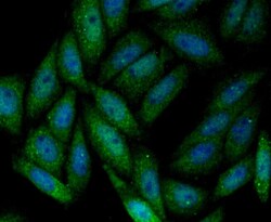
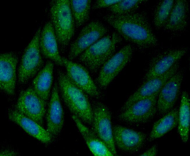
- Immunofluorescent analysis of SENP1 in SiHa cells (green). Samples were fixed in paraformaldehyde and permeabilised with 0.25% Triton X100/PBS, incubated with SENP1 monoclonal antibody (Product # MA5-34767), followed by DAPI (blue).
- Submitted by
- Invitrogen Antibodies (provider)
- Main image

- Experimental details
- Immunofluorescent analysis of SENP1 in SH-SY-5Y cells (green). Samples were fixed in paraformaldehyde and permeabilised with 0.25% Triton X100/PBS, incubated with SENP1 monoclonal antibody (Product # MA5-34767), followed by DAPI (blue).
- Submitted by
- Invitrogen Antibodies (provider)
- Main image
- Experimental details
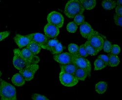
- Immunofluorescent analysis of SENP1 in LOVO cells (green). Samples were fixed in paraformaldehyde and permeabilised with 0.25% Triton X100/PBS, incubated with SENP1 monoclonal antibody (Product # MA5-34767), followed by DAPI (blue).

Supportive validation
- Submitted by
- Invitrogen Antibodies (provider)
- Main image
- Experimental details
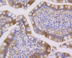
- Immunohistochemistry analysis of SENP1 in paraffin-embedded human small Intestine tissue. Samples were incubated with SENP1 monoclonal antibody (Product # MA5-34767), and followed by hematoxylin.

- Submitted by
- Invitrogen Antibodies (provider)
- Main image

- Experimental details
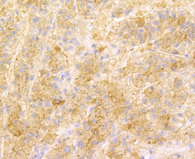
- Immunohistochemistry analysis of SENP1 in paraffin-embedded human liver tissue. Samples were incubated with SENP1 monoclonal antibody (Product # MA5-34767), and followed by hematoxylin.
Supportive validation
- Submitted by
- Invitrogen Antibodies (provider)
- Main image

- Experimental details
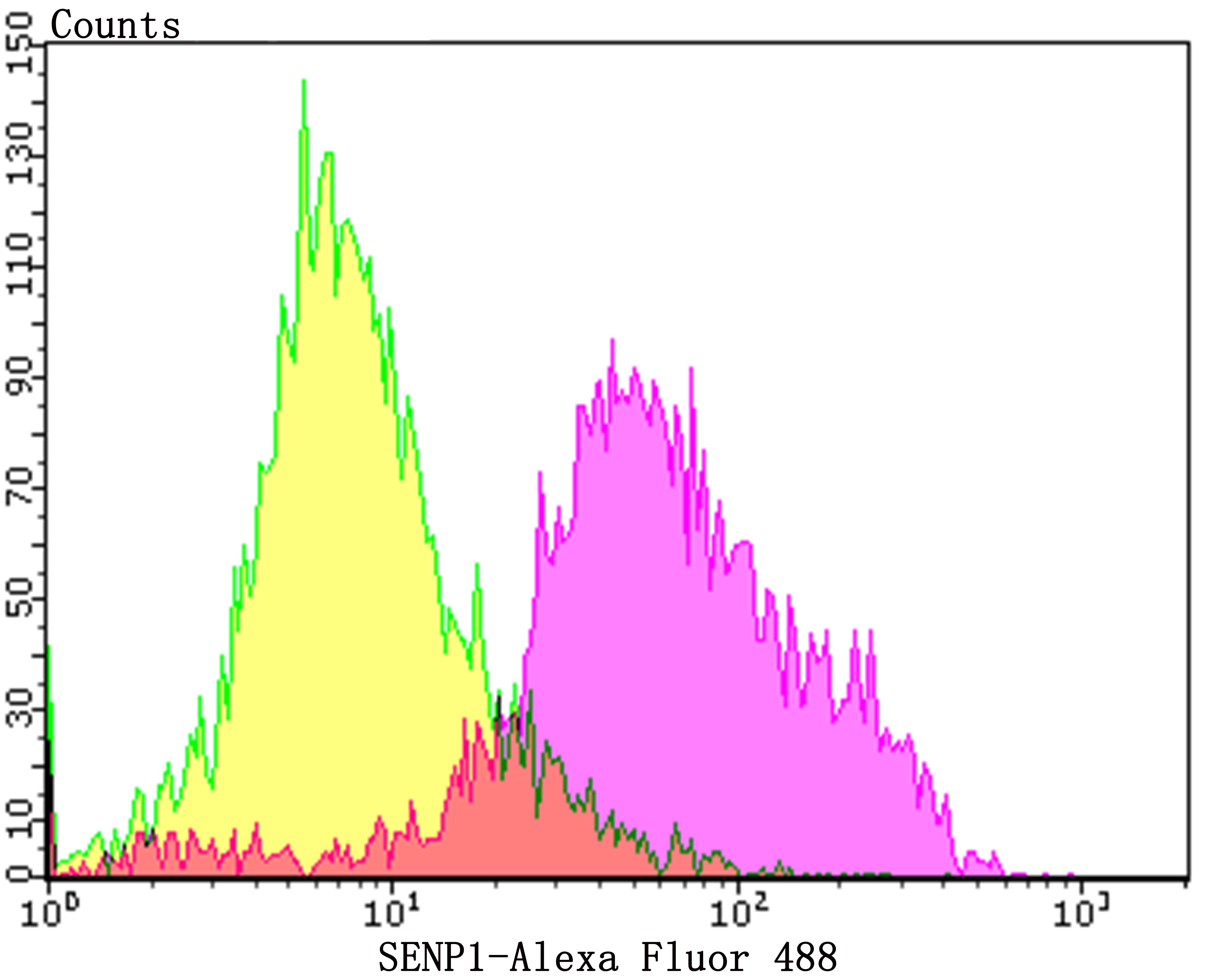
- Flow cytometry of SENP1 in Daudi cells (purple) compared with an unlabelled control (cells without incubation with primary antibody; yellow). Samples were incubated with SENP1 monoclonal antibody (Product # MA5-34767) at a dilution of 1:100, followed by Alexa Fluor 488-conjugated goat anti-rabbit IgG.