 Explore
 Validate
 Learn
Learn Western blot
Antibody data
- Antibody Data
- Antigen structure
- References [1]
- Comments [0]
- Validations
- Western blot [4]
- Immunocytochemistry [1]
- Immunoprecipitation [2]
- Immunohistochemistry [1]
Submit
Validation data
Reference
Comment
Report error
- Product number
- GTX128980 - Provider product page

- Provider
- GeneTex
- Product name
- Visfatin antibody
- Antibody type
- Polyclonal
- Reactivity
- Human, Mouse, Rat
- Host
- Rabbit
Submitted references Renin inhibition improves metabolic syndrome, and reduces angiotensin II levels and oxidative stress in visceral fat tissues in fructose-fed rats.
Chou CL, Lin H, Chen JS, Fang TC
PloS one 2017;12(7):e0180712
PloS one 2017;12(7):e0180712
No comments: Submit comment
Supportive validation
- Submitted by
- GeneTex (provider)
- Main image

- Experimental details
- PBEF antibody detects Visfatin protein by western blot analysis.A. 30 ?g K562 whole cell lysate/extract10% SDS-PAGEVisfatin antibody (GTX128980) dilution: 1:1000 The HRP-conjugated anti-rabbit IgG antibody (GTX213110-01) was used to detect the primary antibody.

- Submitted by
- GeneTex (provider)
- Main image

- Experimental details
- PBEF antibody detects Visfatin protein by western blot analysis.A. 30 ?g 293T whole cell lysate/extract B. 30 ?g A431 whole cell lysate/extract10% SDS-PAGEVisfatin antibody (GTX128980) dilution: 1:1000 The HRP-conjugated anti-rabbit IgG antibody (GTX213110-01) was used to detect the primary antibody.

- Submitted by
- GeneTex (provider)
- Main image

- Experimental details
- Various whole cell extracts (30 ?g) were separated by 10% SDS-PAGE, and the membrane was blotted with Visfatin antibody (GTX128980) diluted at 1:500. The HRP-conjugated anti-rabbit IgG antibody (GTX213110-01) was used to detect the primary antibody.

Supportive validation
- Submitted by
- GeneTex (provider)
- Main image

- Experimental details
- Visfatin antibody detects Visfatin protein at cytoplasm by immunofluorescent analysis.Sample: HeLa cells were fixed in 4% paraformaldehyde at RT for 15 min.Green: Visfatin protein stained by Visfatin antibody (GTX128980) diluted at 1:500.Blue: Hoechst 33342 staining.

Supportive validation
- Submitted by
- GeneTex (provider)
- Main image

- Experimental details
- Immunoprecipitation of Visfatin protein from 293T whole cell extracts using 5 £gg of Visfatin antibody (GTX128980).Western blot analysis was performed using Visfatin antibody (GTX128980).EasyBlot anti-Rabbit IgG (GTX221666-01) was used as a secondary reagent.

Supportive validation
- Submitted by
- GeneTex (provider)
- Main image
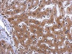
- Experimental details
- PBEF antibody detects Visfatin protein at cytosol on human hepatoma by immunohistochemical analysis. Sample: Paraffin-embedded hepatoma. Visfatin antibody (GTX128980) dilution: 1:500.
