Explore
 Validate
 Learn
Learn Western blot
 ELISA
ELISAAntibody data
- Antibody Data
- Antigen structure
- References [4]
- Comments [0]
- Validations
- Western blot [2]
- Immunohistochemistry [3]
- Other assay [6]
Submit
Validation data
Reference
Comment
Report error
- Product number
- PA5-79470 - Provider product page

- Provider
- Invitrogen Antibodies
- Product name
- IL-17A Polyclonal Antibody
- Antibody type
- Polyclonal
- Antigen
- Recombinant full-length protein
- Description
- Reconstitute with 0.2 mL of distilled water to yield a concentration of 500 µg/mL.
- Reactivity
- Human, Mouse, Rat
- Host
- Rabbit
- Isotype
- IgG
- Vial size
- 100 µg
- Concentration
- 500 µg/mL
- Storage
- -20°C
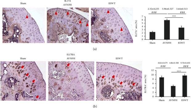
Submitted references Extracorporeal Shockwave Therapy Modulates the Expressions of Proinflammatory Cytokines IL33 and IL17A, and Their Receptors ST2 and IL17RA, within the Articular Cartilage in Early Avascular Necrosis of the Femoral Head in a Rat Model.
Upregulation of Chemokines in the Paraventricular Nucleus of the Hypothalamus in Rats with Stress-Induced Hypertension.
Long-term PM(2.5) exposure increases the risk of non-small cell lung cancer (NSCLC) progression by enhancing interleukin-17a (IL-17a)-regulated proliferation and metastasis.
Neutralization of interleukin-17A alleviates burn-induced intestinal barrier disruption via reducing pro-inflammatory cytokines in a mouse model.
Cheng JH, Jhan SW, Hsu CC, Chiu HW, Hsu SL
Mediators of inflammation 2021;2021:9915877
Mediators of inflammation 2021;2021:9915877
Upregulation of Chemokines in the Paraventricular Nucleus of the Hypothalamus in Rats with Stress-Induced Hypertension.
Wu Q, Chen Y, Zhang W, Song S, Xu Z, Zhang H, Liu L, Sun J
Medical science monitor : international medical journal of experimental and clinical research 2020 Nov 17;26:e926807
Medical science monitor : international medical journal of experimental and clinical research 2020 Nov 17;26:e926807
Long-term PM(2.5) exposure increases the risk of non-small cell lung cancer (NSCLC) progression by enhancing interleukin-17a (IL-17a)-regulated proliferation and metastasis.
Chao X, Yi L, Lan LL, Wei HY, Wei D
Aging 2020 Jun 18;12(12):11579-11602
Aging 2020 Jun 18;12(12):11579-11602
Neutralization of interleukin-17A alleviates burn-induced intestinal barrier disruption via reducing pro-inflammatory cytokines in a mouse model.
Song Y, Li Y, Xiao Y, Hu W, Wang X, Wang P, Zhang X, Yang J, Huang Y, He W, Huang C
Burns & trauma 2019;7:37
Burns & trauma 2019;7:37
No comments: Submit comment
Supportive validation
- Submitted by
- Invitrogen Antibodies (provider)
- Main image

- Experimental details
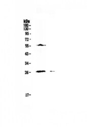
- Western blot analysis of IL-17A in Lane 1: human Jurkat cell lysate using 50 µg (reducing conditions) per well. Electrophoresis was performed on 5-20% SDS-PAGE gel at 70V (Stacking gel) / 90V (Resolving gel) for 2-3 hours and protein was transferred to a nitrocellulose membrane at 150mA for 50-90 minutes. Sample was blocked with 5% Non-fat Milk/TBS for 1.5 hours at room temperature, incubated with IL-17A polyclonal antibody (Product # PA5-79470) at a dilution of 0.5 µg/mL (overnight at 4°C), followed by goat anti-rabbit IgG-HRP secondary antibody at a dilution of 1:10,000. Signal development was performed using a chemiluminescence (ECL) kit.

- Submitted by
- Invitrogen Antibodies (provider)
- Main image
- Experimental details
- Western blot analysis of IL-17A in Lane 1: human Jurkat cell lysate using 50 µg (reducing conditions) per well. Electrophoresis was performed on 5-20% SDS-PAGE gel at 70V (Stacking gel) / 90V (Resolving gel) for 2-3 hours and protein was transferred to a nitrocellulose membrane at 150mA for 50-90 minutes. Sample was blocked with 5% Non-fat Milk/TBS for 1.5 hours at room temperature, incubated with IL-17A polyclonal antibody (Product # PA5-79470) at a dilution of 0.5 µg/mL (overnight at 4°C), followed by goat anti-rabbit IgG-HRP secondary antibody at a dilution of 1:10,000. Signal development was performed using a chemiluminescence (ECL) kit.

Supportive validation
- Submitted by
- Invitrogen Antibodies (provider)
- Main image

- Experimental details
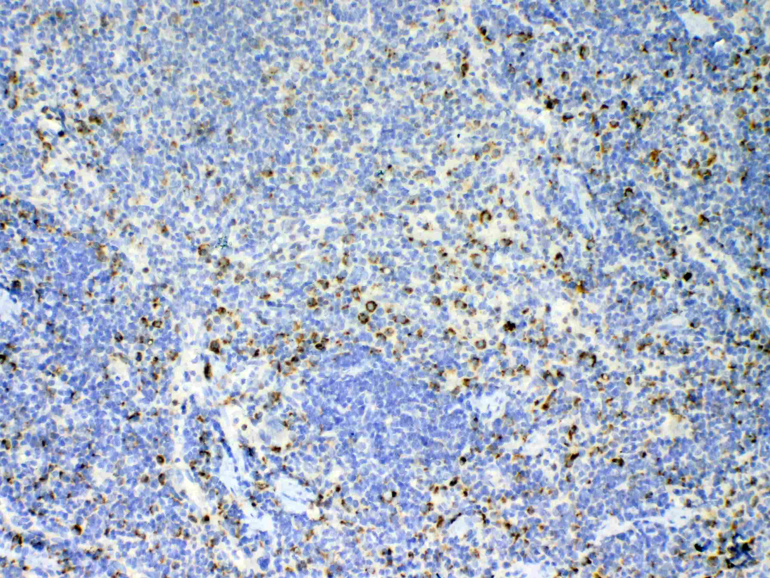
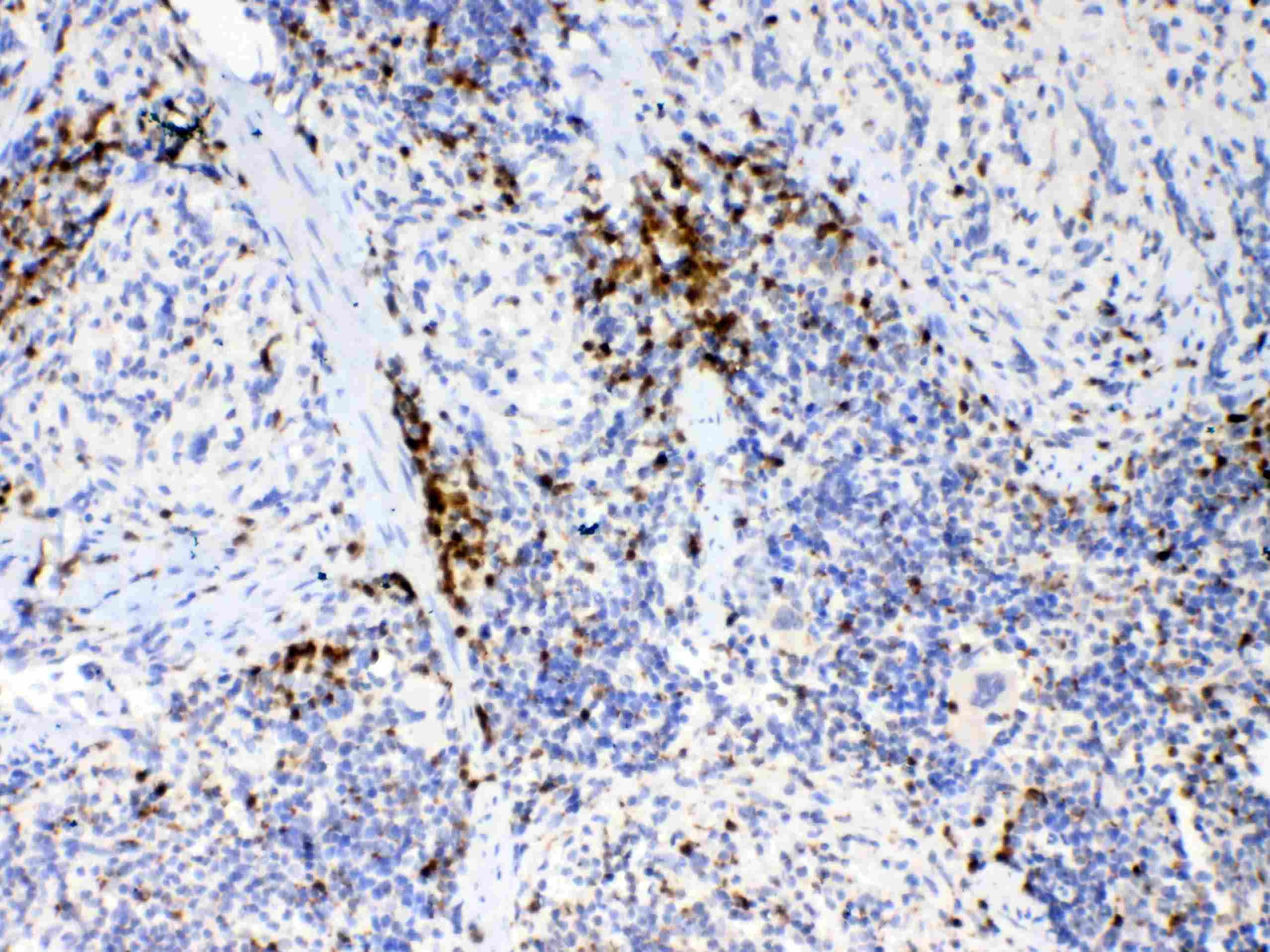
- Immunohistochemistry analysis of IL-17A on paraffin-embedded rat spleen tissue. Antigen retrieval was performed using citrate buffer (pH6, epitope retrieval solution) for 20 mins. Sample was blocked using 10% goat serum, incubated with IL-17A polyclonal antibody (Product# PA5-79470) with a dilution of 1 µg/mL (overnight at 4°C), and followed by biotinylated goat anti-rabbit IgG (30 minutes at 37°C). Development was performed using Streptavidin-Biotin-Complex (SABC) with DAB chromogen method.
- Submitted by
- Invitrogen Antibodies (provider)
- Main image
- Experimental details
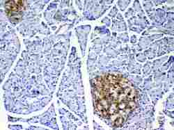
- Immunohistochemistry analysis of IL-17A on paraffin-embedded rat pancreas tissue. Antigen retrieval was performed using citrate buffer (pH6, epitope retrieval solution) for 20 mins. Sample was blocked using 10% goat serum, incubated with IL-17A polyclonal antibody (Product# PA5-79470) with a dilution of 1 µg/mL (overnight at 4°C), and followed by biotinylated goat anti-rabbit IgG (30 minutes at 37°C). Development was performed using Streptavidin-Biotin-Complex (SABC) with DAB chromogen method.

- Submitted by
- Invitrogen Antibodies (provider)
- Main image

- Experimental details
- Immunohistochemistry analysis of IL-17A on paraffin-embedded mouse spleen tissue. Antigen retrieval was performed using citrate buffer (pH6, epitope retrieval solution) for 20 mins. Sample was blocked using 10% goat serum, incubated with IL-17A polyclonal antibody (Product# PA5-79470) with a dilution of 1 µg/mL (overnight at 4°C), and followed by biotinylated goat anti-rabbit IgG (30 minutes at 37°C). Development was performed using Streptavidin-Biotin-Complex (SABC) with DAB chromogen method.
Supportive validation
- Submitted by
- Invitrogen Antibodies (provider)
- Main image
- Experimental details
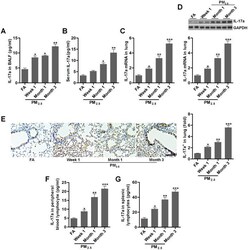
- Figure 3 PM 2.5 enhances IL-17a expression in mice. ( A , B ) IL-17a contents in BALF and serum were measured by ELISA, respectively (n = 8). ( C ) RT-qPCR, ( D ) western blotting and ( E ) IHC analysis of IL-17a in lung tissues of mice challenged with PM 2.5 for the indicated time (n = 6). Scale bar, 100 mum. ( F , G ) IL-17a levels in the peripheral blood lymphocytes and in splenic lymphocytes were calculated using ELISA analysis (n = 8). All data are expressed as mean +- SEM. * p

- Submitted by
- Invitrogen Antibodies (provider)
- Main image

- Experimental details
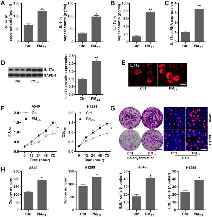
- Figure 5 PM 2.5 elevates the proliferation in NSCLC cells. ( A - E ) Th17 cells were treated with 100 mug/cm 2 of PM 2.5 for 24 h, and then all cells and supernatants were collected for the analysis. ( A ) TNF-alpha and IL-6 levels in the supernatants were measured using ELISA. ( B ) IL-17a contents in the collected supernatants were calculated using ELISA. ( C ) RT-qPCR and ( D ) western blot analysis were used to measure IL-17a expression levels in the harvested cells. ( E ) IF staining of IL-17a in the harvested cells. Scale bar, 20 mum. ( F ) Th17 cells were treated with 100 mug/cm 2 of PM 2.5 for 24 h, and then the conditional medium was collected, and mixed with fresh RPMI1640 absolute medium at 1:3. The composed culture medium was exposed to A549 and H1350 cells for 12, 24, 48 or 72 h. Then, the NSCLC cells were collected for cell proliferation analysis using CCK-8 analysis. ( G , H ) Th17 cells were incubated with 100 mug/cm 2 of PM 2.5 for 24 h, and then the conditional medium was collected, and mixed with fresh RPMI1640 absolute medium at 1:3. Then, the composed culture medium was subjected to A549 and H1350 cells for another 24 h. Subsequently, all cells were harvested to assess the cell proliferation using colony formation and EdU assays. Scale bar, 100 mum. Quantification of colony formation assay and EdU was exhibited. All data are expressed as mean +- SEM. * p
- Submitted by
- Invitrogen Antibodies (provider)
- Main image

- Experimental details
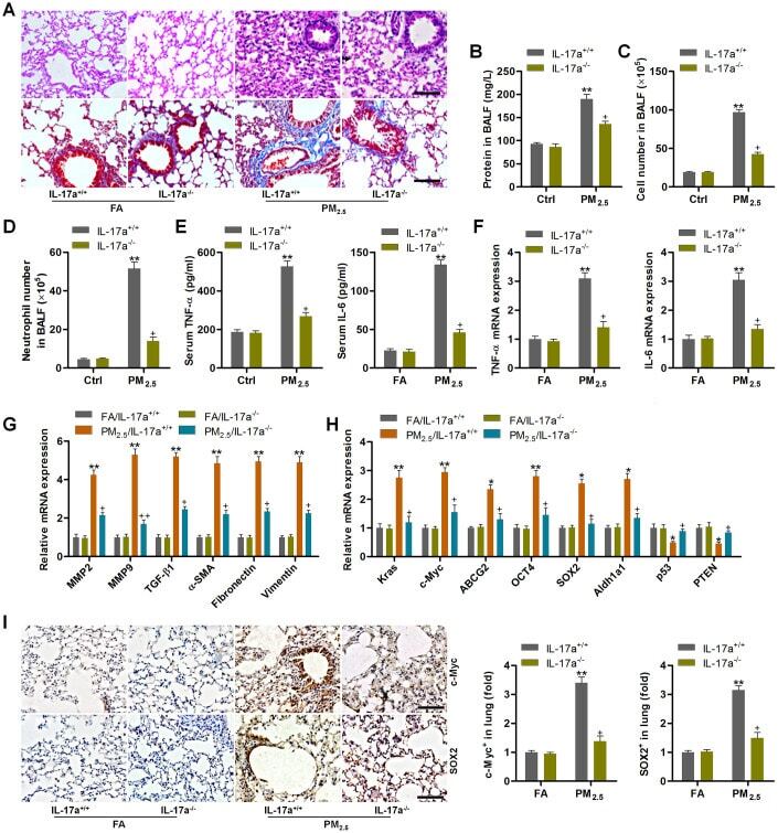
- Figure 8 IL-17a knockout alleviates pulmonary injury and cancer stem cell properties in mice following PM 2.5 exposure for 3 months. ( A ) H&E staining (up panel) and Masson trichrome staining (down panel) of lung sections from IL-17a +/+ and IL-17a -/- mice challenged with or without PM 2.5 for 3 months (n = 6). Scale bar, 100 mum. ( B ) Protein levels in BALF were measured (n = 8). ( C ) The total number of cells in BALF was assessed (n = 8). ( D ) The number of neutrophils in BALF was measured (n = 8). ( E ) Serum TNF-alpha and IL-6 levels in mice were measured by ELISA (n = 8). ( F ) TNF-alpha and IL-6 mRNA levels in the pulmonary samples were determined using RT-qPCR analysis (n = 4). ( G ) Fibrosis-associated genes as shown were tested using RT-qPCR analysis (n = 4). ( H ) Genes associated with lung cancer progression were calculated using RT-qPCR analysis (n = 4). ( I ) IHC staining of c-Myc and SOX2 in pulmonary sections from the indicated groups of mice (n = 6). Scale bar, 100 mum. All data are expressed as mean +- SEM. * p
- Submitted by
- Invitrogen Antibodies (provider)
- Main image

- Experimental details
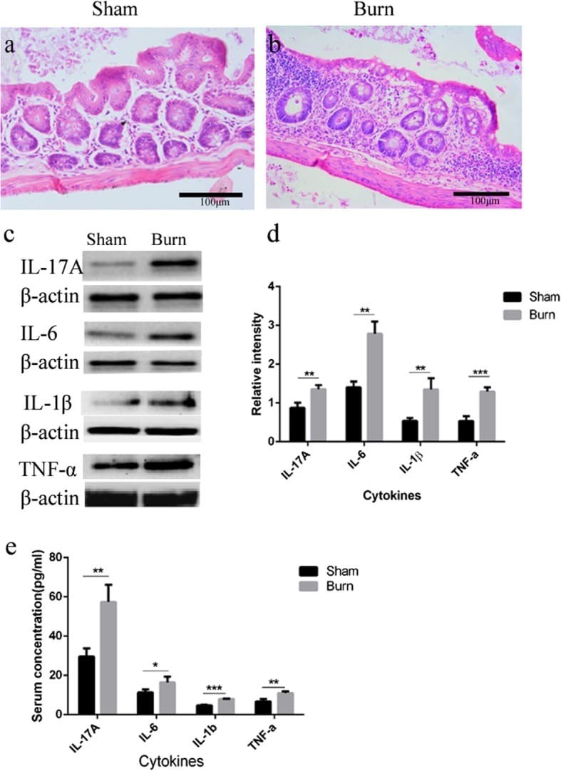
- Fig. 2 Epithelial inflammatory cell infiltration, and elevated pro-inflammatory cytokine expression induced by burn injury. Ileal sections were stained with hematoxylin and eosin (H&E), and extensive microscopic inflammatory cell infiltration in burn group compared with the sham group was observed ( a , b , 200x). Cytokine protein levels in ileum of both groups were determined by Western blotting ( c ), and a summary of cytokines blots were presented as the ratio of cytokines:beta-actin densities ( d ). Cytokine protein levels in serum of both groups were determined by enzyme-linked immunosorbent assay (ELISA), and normalized to pg/ml of total serum volume ( e ). n = 5 per group. * p < 0.05, ** p < 0.01, *** p < 0.001. IL Interleukin, TNF-alpha Tumor necrosis factor-alpha
- Submitted by
- Invitrogen Antibodies (provider)
- Main image

- Experimental details
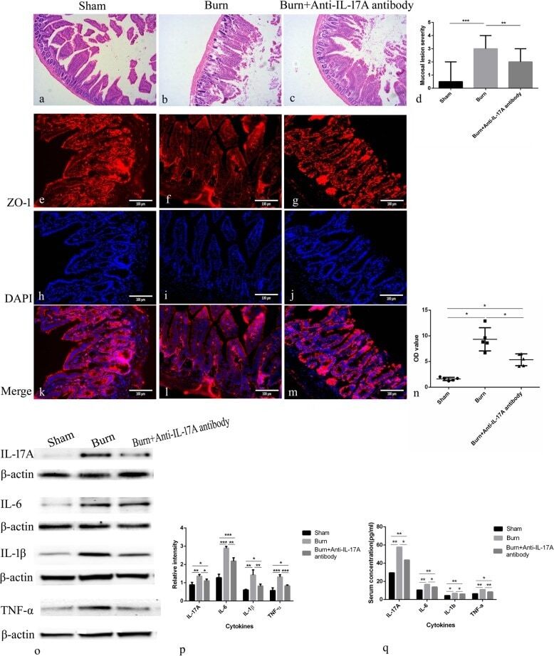
- Fig. 3 Interleukin (IL)-17A neutralization attenuated intestinal pathological changes and zonula occludens-1 (ZO-1) alteration, suppressed intestinal permeability elevation, and inhibited increases of cytokines. Ileal sections were stained with hematoxylin and eosin (H&E) ( a , b , c 200x), and mucosa lensions were scored in each group( d ). Representative immunofluorescent images depicting membrane localization of ZO-1 in sham group ( e - g ), burn group ( h - j ), and burn+anti-IL-17A antibody group ( l - m ), red staining indicates ZO-1, and blue staining indicates nuclei (400x). Intestinal permeability was measured by fluorescein isothiocyanate (FITC)-dextran levels and is expressed as optical density (OD) value (mean +- standard error of the mean (SEM)) in per group ( n ). Cytokine protein levels in ileum of each group were determined by Western blotting ( o ), and summary of cytokines blots is presented as the ratio of cytokines: beta-actin densities ( p ). Cytokine protein levels in serum of each group were determined by enzyme-linked immunosorbent assay (ELISA), and normalized to pg/ml of total serum volume ( q ). n = 5 per group. * p < 0.05, ** p < 0.01, *** p < 0.001. DAPI 4',6-diamidino-2-phenylindole
- Submitted by
- Invitrogen Antibodies (provider)
- Main image
- Experimental details
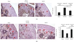
- Figure 6 Immunohistochemical analysis for (a) IL17, (b) IL17RA in the articular cartilage of the left femur head (right), and the level of expression was measured after treatment (left). The expression of IL17A was major in superfacial zone and proliferation zone (arrowhead), while IL17RA was major expressed in the hypertrophic chondrocytes of the calcified cartilage zone (arrow). *** P < 0.001 as compared with the ESWT group and ### P < 0.001 as compared with the AVNFH group. The scale bar was 50 mu m. N = 8 for all groups.