 Explore
 Validate
 Learn
Learn Western blot
Antibody data
- Antibody Data
- Antigen structure
- References [1]
- Comments [0]
- Validations
- Western blot [3]
- Other assay [1]
Submit
Validation data
Reference
Comment
Report error
- Product number
- PA5-50485 - Provider product page

- Provider
- Invitrogen Antibodies
- Product name
- SLC7A5 Polyclonal Antibody
- Antibody type
- Polyclonal
- Antigen
- Synthetic peptide
- Description
- The antibody detects endogenous levels of total CD98 / SLC3A2 protein.
- Reactivity
- Human
- Host
- Rabbit
- Isotype
- IgG
- Vial size
- 100 µL
- Concentration
- 0.4 mg/mL
- Storage
- -20°C
Submitted references A novel therapeutic approach for anaplastic thyroid cancer through inhibition of LAT1.
Enomoto K, Sato F, Tamagawa S, Gunduz M, Onoda N, Uchino S, Muragaki Y, Hotomi M
Scientific reports 2019 Oct 10;9(1):14616
Scientific reports 2019 Oct 10;9(1):14616
No comments: Submit comment
Supportive validation
- Submitted by
- Invitrogen Antibodies (provider)
- Main image

- Experimental details
- Western blot analysis of Slc7a5 was performed by loading (from left to right): Mouse liver tissue lysates (40µg) on to a 8% SDS-PAGE gel. Proteins were transferred to a membrane and the membrane was probed with a Slc7a5 antibody (Product # PA5-50485) at a 1/200 dilution for 2 minutes, followed by a secondary antibody at 1/8000.

- Submitted by
- Invitrogen Antibodies (provider)
- Main image
- Experimental details
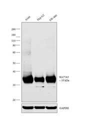
- Western blot analysis was performed on membrane enriched extracts (30 µg lysate) of A549 (Lane 1), Hep G2 (Lane 2) and SW-480 (Lane 3). The blot was probed with Anti-SLC7A5 Polyclonal Antibody (Product # PA5-50485, 1:500 dilution) and detected by chemiluminescence using Goat anti-Rabbit IgG (H+L) Superclonal™ Secondary Antibody, HRP conjugate (Product # A27036, 0.25 µg/ml, 1:4000 dilution). A 35 kDa band corresponding to SLC7A5 was observed across cell lines tested.

- Submitted by
- Invitrogen Antibodies (provider)
- Main image

- Experimental details
- Knockdown of SLC7A5 was achieved by transfecting A549 cells with SLC7A5 specific siRNAs (Silencer® select Product # s15655, s15653). Western blot analysis (Fig. a) was performed using membrane enriched extracts from the SLC7A5 knockdown cells (lane 3), non-specific scrambled siRNA transfected cells (lane 2) and untransfected cells (lane 1). The blot was probed with SLC7A5 Polyclonal Antibody (Product # PA5-50485, 1:500 dilution) and Goat anti-Rabbit IgG (H+L) Superclonal™ Secondary Antibody, HRP conjugate (Product # A27036, 0.25µg/ml, 1:4000 dilution). Densitometric analysis of this western blot is shown in histogram (Fig. b). Decrease in signal upon siRNA mediated knock down confirms that antibody is specific to SLC7A5.

Supportive validation
- Submitted by
- Invitrogen Antibodies (provider)
- Main image

- Experimental details
- Figure 2 Detection of LAT1 and 4F2hc by western blotting and immunofluorescence analyses in 8505C, OCUT-2, and OCUT-6 human ATC cells. ( A ) Expression of LAT1 and 4F2hc levels in the indicated cells detected by western blotting. ( B ) The densitometry value of LAT1 was normalized alpha-tubulin. ( C ) The densitometry value of 4F2hc was normalized alpha-tubulin. ( D ) DAPI (4,6-diamidino-2-phenylindole is a blue fluorescent stain specific for DNA) nuclear staining was shown in blue. Both LAT1 (green) and 4F2hc immunoreactivities (red) were detected on the plasma membrane of 8505C, OCUT-2, or OCUT-6 cells. LAT1 (green) and 4F2hc immunostaining (red), and DAPI (blue) were merged and shown in 2D, indicating the coexistence of LAT1 with 4F2hc in the plasma membrane of 8505C, OCUT-2, or OCUT-6 cells, respectively.
