 Explore
 Validate
 Learn
Learn Western blot
 Immunoprecipitation
Antibody data
- Antibody Data
- Antigen structure
- References [0]
- Comments [0]
- Validations
- Western blot [4]
- Immunocytochemistry [2]
Submit
Validation data
Reference
Comment
Report error
- Product number
- PA5-17017 - Provider product page

- Provider
- Invitrogen Antibodies
- Product name
- RRAGC Polyclonal Antibody
- Antibody type
- Polyclonal
- Antigen
- Synthetic peptide
- Description
- It is not recommended to aliquot this antibody.
- Reactivity
- Human, Mouse, Rat
- Host
- Rabbit
- Isotype
- IgG
- Vial size
- 100 µL
- Concentration
- 19 µg/mL
- Storage
- -20°C
No comments: Submit comment
Supportive validation
- Submitted by
- Invitrogen Antibodies (provider)
- Main image

- Experimental details
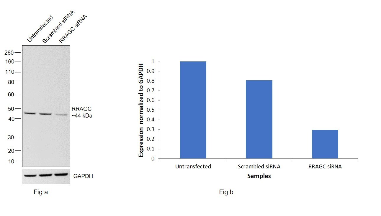
- Knockdown of RRAGC was achieved by transfecting A-431 with RRAGC specific siRNAs (Silencer® select Product # s224599, s224598). Western blot analysis (Fig. a) was performed using Whole cell extracts from the RRAGC knockdown cells (lane 3), non-targeting scrambled siRNA transfected cells (lane 2), and untransfected cells (lane 1). The blot was probed with RRAGC Polyclonal Antibody (Product # PA5-17017, 1:1000) and Goat anti-Rabbit IgG (H+L) Superclonal™ Recombinant Secondary Antibody, HRP (Product # A27036, 1:4000). Densitometric analysis of this western blot is shown in the histogram (Fig. b). A decrease in signal upon siRNA mediated knockdown confirms that antibody is specific to RRAGC.
- Submitted by
- Invitrogen Antibodies (provider)
- Main image

- Experimental details
- Western blot was performed using Anti-RRAGC Polyclonal Antibody(Product # PA5-17017) and a 44kDa band corresponding to RRAGC was observed across the cell lines and tissues tested. Whole cell extracts (30 µg lysate) of HEK-293 (Lane 1), A-431 (Lane 2), BeWo (Lane 3), PANC-1 (Lane 4), Raji (Lane 5), Mouse Lung (Lane 6) and Rat Lung (Lane 7) were electrophoresed using NuPAGE™ 4-12% Bis-Tris Protein Gel (Product # NP0322BOX). Resolved proteins were then transferred onto a Nitrocellulose membrane (Product # IB23001) by iBlot® 2 Dry Blotting System (Product # IB21001). The blot was probed with the primary antibody (1:1000 dilution) and detected by chemiluminescence with Goat anti-Rabbit IgG (H+L) Superclonal™ Recombinant Secondary Antibody, HRP (Product # A27036, 1:4000 dilution) using the iBright FL 1000 (Product # A32752). Chemiluminescent detection was performed using Novex® ECL Chemiluminescent Substrate Reagent Kit (Product # WP20005).

- Submitted by
- Invitrogen Antibodies (provider)
- Main image
- Experimental details
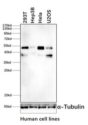
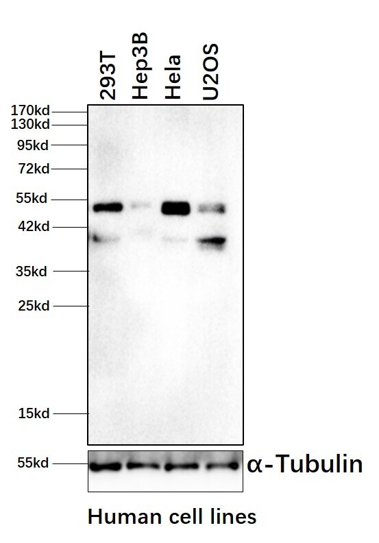
- Western blot analysis of RRAGC was performed by loading whole-cell lysates of 293T (lane 1), Hep3B (lane 2), HeLa (lane 3) and U2OS (lane 4). RRAGC was detected at approximately 50kDa using anti-RRAGC Polyclonal Antibody (Product # PA5-17017) at a dilution of 1:1000 in Blocking Buffer overnight at 4°C on a rocking platform, followed by a 1-hour room-temperature incubation using a donkey anti-rabbit IgG antibody at a dilution of 1:5000. Data courtesy of Thermo Scientific KOL Program.
- Submitted by
- Invitrogen Antibodies (provider)
- Main image
- Experimental details
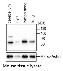
- Western blot analysis of RRAGC was performed by loading whole-cell lysates of mouse tissue samples. RRAGC was detected at approximately 50kDa by using a RRAGC polyclonal Antibody (Product # PA5-17017) at a dilution of 1:1000 in Blocking Buffer overnight at 4°C on a rocking platform, followed by a 1-hour room-temperature incubation of a donkey anti-rabbit IgG antibody at a dilution of 1:5000. Data courtesy of Thermo Scientific KOL Program.

Supportive validation
- Submitted by
- Invitrogen Antibodies (provider)
- Main image
- Experimental details
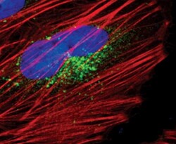
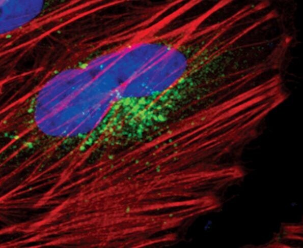
- Immunofluorescent analysis of RagC in HeLa cells using a RagC polyclonal antibody (Product # PA5-17017) (green). Actin filaments are labeled with a fluorescent red phalloidin. DNA is labeled using a fluorescent blue dye.
- Submitted by
- Invitrogen Antibodies (provider)
- Main image
- Experimental details
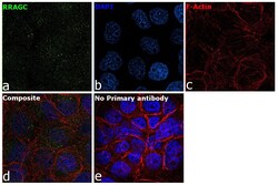
- Immunofluorescence analysis of RRAGC was performed using 70% confluent log phase A-431 cells. The cells were fixed with 4% paraformaldehyde for 10 minutes, permeabilized with 0.1% Triton™ X-100 for 15 minutes, and blocked with 2% BSA for 45 minutes at room temperature. The cells were labeled with RRAGC Polyclonal Antibody (Product # PA5-17017) at 1:200 in 0.1% BSA, incubated at 4 degree celsius overnight and then labeled with Donkey anti-Rabbit IgG (H+L) Highly Cross-Adsorbed Secondary Antibody, Alexa Fluor Plus 488 (Product # A32790), (1:2000 dilution), for 45 minutes at room temperature (Panel a: Green). Nuclei (Panel b:Blue) were stained with ProLong™ Diamond Antifade Mountant with DAPI (Product # P36962). F-actin (Panel c: Red) was stained with Rhodamine Phalloidin (Product # R415, 1:300). Panel d represents the merged image showing Cytoplasmic localization. Panel e represents control cells with no primary antibody to assess background. The images were captured at 60X magnification.
