Explore
 Validate
 Learn
Learn Western blot
Antibody data
- Antibody Data
- Antigen structure
- References [9]
- Comments [0]
- Validations
- Western blot [1]
Submit
Validation data
Reference
Comment
Report error
- Product number
- MAB868 - Provider product page

- Provider
- R&D Systems
- Product name
- Human APAF-1 Antibody
- Antibody type
- Monoclonal
- Description
- Protein A or G purified from hybridoma culture supernatant. Detects human APAF-1 in Western blots.
- Reactivity
- Human
- Host
- Mouse
- Conjugate
- Unconjugated
- Antigen sequence
AAC51678- Isotype
- IgG
- Antibody clone number
- 94408
- Vial size
- 100 ug
- Concentration
- LYOPH
- Storage
- Use a manual defrost freezer and avoid repeated freeze-thaw cycles. 12 months from date of receipt, -20 to -70 °C as supplied. 1 month, 2 to 8 °C under sterile conditions after reconstitution. 6 months, -20 to -70 °C under sterile conditions after reconstitution.
Submitted references Delayed apoptosis allows islet β-cells to implement an autophagic mechanism to promote cell survival.
Ash2L enables P53-dependent apoptosis by favoring stable transcription pre-initiation complex formation on its pro-apoptotic target promoters.
A threshold mechanism mediates p53 cell fate decision between growth arrest and apoptosis.
Alteration of the nuclear pore complex in Ca(2+)-mediated cell death.
Intracellular K(+) inhibits apoptosis by suppressing the Apaf-1 apoptosome formation and subsequent downstream pathways but not cytochrome c release.
Fas-mediated apoptosome formation is dependent on reactive oxygen species derived from mitochondrial permeability transition in Jurkat cells.
Fas-mediated apoptosome formation is dependent on reactive oxygen species derived from mitochondrial permeability transition in Jurkat cells.
The role of mitochondria, cytochrome c and caspase-9 in embryonic lens fibre cell denucleation.
The role of mitochondria, cytochrome c and caspase-9 in embryonic lens fibre cell denucleation.
Hayes HL, Peterson BS, Haldeman JM, Newgard CB, Hohmeier HE, Stephens SB
PloS one 2017;12(2):e0172567
PloS one 2017;12(2):e0172567
Ash2L enables P53-dependent apoptosis by favoring stable transcription pre-initiation complex formation on its pro-apoptotic target promoters.
Mungamuri SK, Wang S, Manfredi JJ, Gu W, Aaronson SA
Oncogene 2015 May 7;34(19):2461-70
Oncogene 2015 May 7;34(19):2461-70
A threshold mechanism mediates p53 cell fate decision between growth arrest and apoptosis.
Kracikova M, Akiri G, George A, Sachidanandam R, Aaronson SA
Cell death and differentiation 2013 Apr;20(4):576-88
Cell death and differentiation 2013 Apr;20(4):576-88
Alteration of the nuclear pore complex in Ca(2+)-mediated cell death.
Bano D, Dinsdale D, Cabrera-Socorro A, Maida S, Lambacher N, McColl B, Ferrando-May E, Hengartner MO, Nicotera P
Cell death and differentiation 2010 Jan;17(1):119-33
Cell death and differentiation 2010 Jan;17(1):119-33
Intracellular K(+) inhibits apoptosis by suppressing the Apaf-1 apoptosome formation and subsequent downstream pathways but not cytochrome c release.
Karki P, Seong C, Kim JE, Hur K, Shin SY, Lee JS, Cho B, Park IS
Cell death and differentiation 2007 Dec;14(12):2068-75
Cell death and differentiation 2007 Dec;14(12):2068-75
Fas-mediated apoptosome formation is dependent on reactive oxygen species derived from mitochondrial permeability transition in Jurkat cells.
Sato T, Machida T, Takahashi S, Iyama S, Sato Y, Kuribayashi K, Takada K, Oku T, Kawano Y, Okamoto T, Takimoto R, Matsunaga T, Takayama T, Takahashi M, Kato J, Niitsu Y
Journal of immunology (Baltimore, Md. : 1950) 2004 Jul 1;173(1):285-96
Journal of immunology (Baltimore, Md. : 1950) 2004 Jul 1;173(1):285-96
The role of mitochondria, cytochrome c and caspase-9 in embryonic lens fibre cell denucleation.
Sanders EJ, Parker E
Journal of anatomy 2002 Aug;201(2):121-35
Journal of anatomy 2002 Aug;201(2):121-35
The role of mitochondria, cytochrome c and caspase-9 in embryonic lens fibre cell denucleation.
Sanders EJ, Parker E
Journal of anatomy 2002 Aug;201(2):121-35
Journal of anatomy 2002 Aug;201(2):121-35
No comments: Submit comment
Supportive validation
- Submitted by
- R&D Systems (provider)
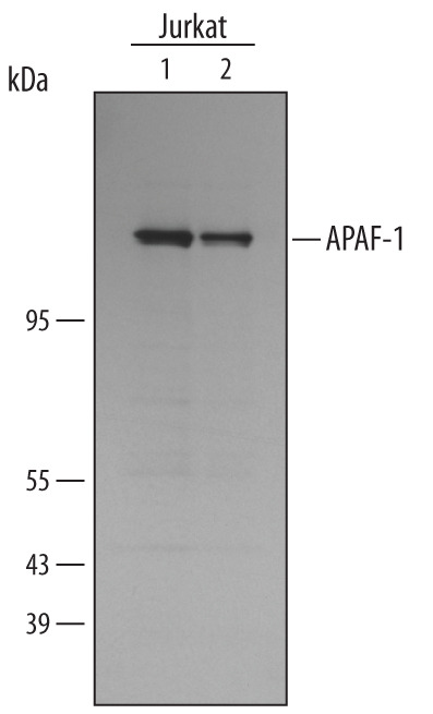
- Main image
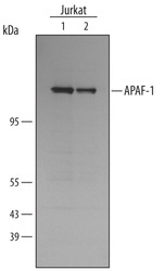
- Experimental details
- Detection of Human APAF-1 by Western Blot. Western blot shows lysates of Jurkat human acute T cell leukemia cell line. Gels were loaded with 2 x 10⁵ cells (lane 1) and 7 x 10⁴ cells (lane 2). PVDF membrane was probed with 1 µg/mL Mouse Anti-Human APAF-1 Monoclonal Antibody (Catalog # MAB868) followed by HRP-conjugated Anti-Mouse IgG Secondary Antibody (Catalog # HAF007). A specific band for APAF-1 was detected at approximately 135 kDa (as indicated). This experiment was conducted under reducing conditions and using Immunoblot Buffer Group 4.