 Explore
 Validate
 Learn
Learn Western blot
 Immunoprecipitation
Antibody data
- Antibody Data
- Antigen structure
- References [0]
- Comments [0]
- Validations
- Western blot [4]
Submit
Validation data
Reference
Comment
Report error
- Product number
- MA1-23191 - Provider product page

- Provider
- Invitrogen Antibodies
- Product name
- DNA Ligase III Monoclonal Antibody (1F3)
- Antibody type
- Monoclonal
- Antigen
- Recombinant full-length protein
- Description
- Recommended positive controls: 293T, A431, HeLa, HepG2. The predicted molecular weight of MA1-23191 is 100 kDa.
- Reactivity
- Human, Mouse, Chicken/Avian
- Host
- Mouse
- Isotype
- IgG
- Antibody clone number
- 1F3
- Vial size
- 100 µL
- Concentration
- 1 mg/mL
- Storage
- Store at 4°C short term. For long term storage, store at -20°C, avoiding freeze/thaw cycles.
No comments: Submit comment
Supportive validation
- Submitted by
- Invitrogen Antibodies (provider)
- Main image

- Experimental details
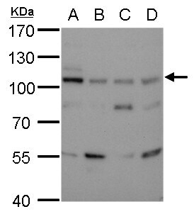
- DNA ligase III detects DNA Ligase III Monoclonal Antibody (1F3) protein by western blot analysis. A. 30 µg 293T whole cell lysate/extract. B. 30 µg A431 whole cell lysate/extract. C. 30 µg HeLa whole cell lysate/extract. D. 30 µg HepG2 whole cell lysate/extract.7.5% SDS-PAGE. DNA ligase III DNA Ligase III Monoclonal Antibody (1F3) (Product # MA1-23191) dilution: 1:500. The HRP-conjugated anti-mouse IgG antibody was used to detect the primary antibody.
- Submitted by
- Invitrogen Antibodies (provider)
- Main image
- Experimental details
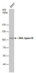
- DNA Ligase III Polyclonal Antibody detects DNA ligase III protein by western blot analysis. Whole cell extracts (30 µg) was separated by 7.5% SDS-PAGE, and the membrane was blotted with DNA Ligase III Polyclonal Antibody DNA Ligase III Monoclonal Antibody (1F3) (Product # MA1-23191) diluted by 1:500. The HRP-conjugated anti-mouse IgG antibody was used to detect the primary antibody.

- Submitted by
- Invitrogen Antibodies (provider)
- Main image

- Experimental details
- Knockdown of DNA Ligase III was achieved by transfecting HeLa cells with LIG3 specific siRNAs (Silencer® select Product # S8177, S8178). Western blot analysis (Fig. a) was performed using whole cell extracts from the Cyclin D1 knockdown cells (lane 3), non-specific scrambled siRNA transfected cells (lane 2) and untransfected cells (lane 1). The blot was probed with DNA Ligase III Monoclonal Antibody (1F3) (Product # MA1-23191, 1:1000 dilution) and Goat anti-Mouse IgG (H+L) Superclonal™ Secondary Antibody, HRP (Product # A28177, 1:4000 dilution). Densitometric analysis of this western blot is shown in histogram (Fig. b). Decrease in signal upon siRNA mediated knock down confirms that antibody is specific to DNA Ligase III.

- Submitted by
- Invitrogen Antibodies (provider)
- Main image

- Experimental details
- Western blot was performed using DNA Ligase III Monoclonal Antibody (1F3) (Product # PA1-23191) and a 100kDa band corresponding to DNA Ligase III was observed across all the cell lines tested with uncharacterized non specific band (*). Whole cell extracts (30 µg lysate) of K-562 (Lane 1), Jurkat (Lane 2), U2 OS (Lane 3), HeLa (Lane 4) were electrophoresed using NuPAGE™ 4-12% Bis-Tris Protein Gel (Product # NP0321BOX). Resolved proteins were then transferred onto a Nitrocellulose membrane (Product # IB23001) by iBlot® 2 Dry Blotting System (Product # IB21001). The blot was probed with the primary antibody (1:1000 dilution) and detected by chemiluminescence with Goat anti-Mouse IgG (H+L), Superclonal™ Recombinant Secondary Antibody, HRP (Product # A28177, 1:4000 dilution) using the iBright FL 1000 (Product # A32752). Chemiluminescent detection was performed using Novex® ECL Chemiluminescent Substrate Reagent Kit (Product # WP20005).
