 Explore
 Validate
 Learn
Learn Western blot
Antibody data
- Antibody Data
- Antigen structure
- References [0]
- Comments [0]
- Validations
- Western blot [3]
- ELISA [1]
- Immunocytochemistry [1]
- Immunohistochemistry [5]
Submit
Validation data
Reference
Comment
Report error
- Product number
- TA590235 - Provider product page

- Provider
- OriGene
- Product name
- Rabbit Polyclonal ABCF2 Antibody
- Antibody type
- Polyclonal
- Description
- Rabbit Polyclonal ABCF2 Antibody
- Host
- Rabbit
- Conjugate
- Unconjugated
- Epitope
- ABCF2
- Isotype
- IgG
- Antibody clone number
- NULL
- Vial size
- 100 µg
- Concentration
- 0.72117 mg/ml
No comments: Submit comment
Supportive validation
- Submitted by
- OriGene (provider)
- Main image
- Experimental details
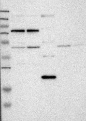
- Lane 1: Marker [kDa] 250, 130, 95, 72, 55, 36, 28, 17, 11; Lane 2: RT-4; Lane 3: U-251 MG; Lane 4: Human Plasma; Lane 5: Liver; Lane 6: TonsilThis validation was performed by Protein Atlas and the presentation of data is for informational purposes only.
- Validation comment
- WB

- Submitted by
- OriGene (provider)
- Main image
- Experimental details
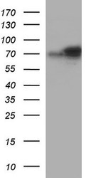
- HEK293T cells were transfected with the pCMV6-ENTRY control (Left lane) or pCMV6-ENTRY ABCF2 (RC200323, Right lane) cDNA for 48 hrs and lysed. Equivalent amounts of cell lysates (5 ug per lane) were separated by SDS-PAGE and immunoblotted with anti-ABCF2.
- Validation comment
- WB

- Submitted by
- OriGene (provider)
- Main image

- Experimental details
- Western blot analysis of extracts (35ug) from 9 different cell lines by using anti-ABCF2 polyclonal antibody (HepG2: human; HeLa: human; SVT2: mouse; A549: human; COS7: monkey; Jurkat: human; MDCK: canine; PC12: rat; MCF7: human).
- Validation comment
- WB

Supportive validation
- Submitted by
- OriGene (provider)
- Main image
- Experimental details
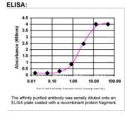
- ELISA: ABCF2 Antibody
- Validation comment
- ELISA

Supportive validation
- Submitted by
- OriGene (provider)
- Main image
- Experimental details
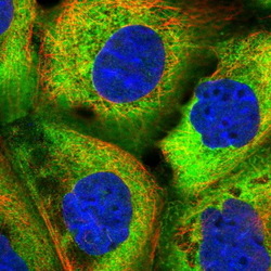
- Immunofluorescent staining of human cell line A-431 shows positivity in cytoplasm.This validation was performed by Protein Atlas and the presentation of data is for informational purposes only.
- Validation comment
- IF

Supportive validation
- Submitted by
- OriGene (provider)
- Main image
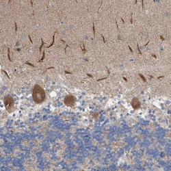
- Experimental details
- Immunohistochemical staining of human cerebellum shows distinct cytoplasmic positivity in Purkinje cells.This validation was performed by Protein Atlas and the presentation of data is for informational purposes only.
- Validation comment
- IHC

- Submitted by
- OriGene (provider)
- Main image
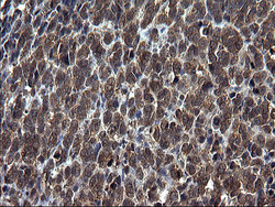
- Experimental details
- Immunohistochemical staining of paraffin-embedded Adenocarcinoma of Human colon tissue using anti-ABCF2 rabbit polyclonal antibody. (TA590235)
- Validation comment
- IHC
- Submitted by
- OriGene (provider)
- Main image

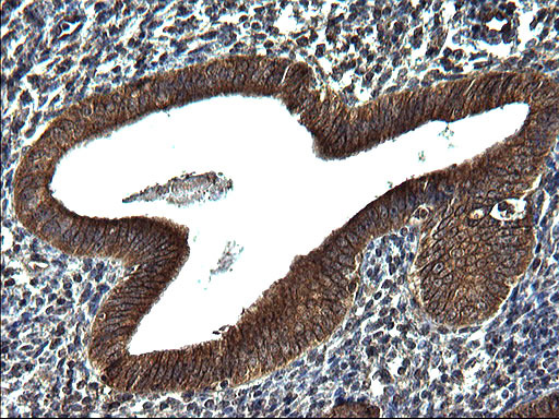
- Experimental details
- Immunohistochemical staining of paraffin-embedded Human endometrium tissue using anti-ABCF2 rabbit polyclonal antibody. (TA590235)
- Validation comment
- IHC
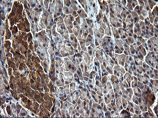
- Submitted by
- OriGene (provider)
- Main image
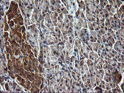
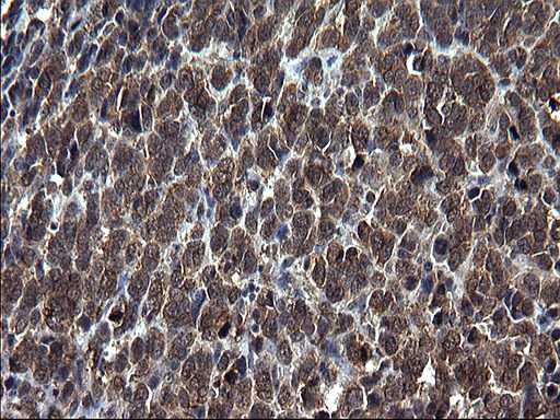
- Experimental details
- Immunohistochemical staining of paraffin-embedded Human pancreas tissue using anti-ABCF2 rabbit polyclonal antibody. (TA590235)
- Validation comment
- IHC
- Submitted by
- OriGene (provider)
- Main image
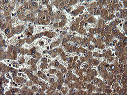
- Experimental details
- Immunohistochemical staining of paraffin-embedded Human liver tissue using anti-ABCF2 rabbit polyclonal antibody. (TA590235)
- Validation comment
- IHC
