 Explore
 Validate
 Learn
Learn Western blot
 ELISA
ELISAAntibody data
- Antibody Data
- Antigen structure
- References [1]
- Comments [0]
- Validations
- Western blot [2]
- Immunohistochemistry [1]
- Flow cytometry [1]
Submit
Validation data
Reference
Comment
Report error
- Product number
- OAAB03804 - Provider product page

- Provider
- Aviva Systems Biology
- Product name
- HPRT1 antibody - C-terminal region
- Antibody type
- Polyclonal
- Reactivity
- Human
- Host
- Rabbit
- Vial size
- 400ul
- Storage
- Maintain refrigerated at 2-8 deg C for up to 6 months. For long term storage store at -20 deg C in small aliquots to prevent freeze-thaw cycles.
Submitted references Loss of dopamine phenotype among midbrain neurons in Lesch-Nyhan disease.
Göttle M, Prudente CN, Fu R, Sutcliffe D, Pang H, Cooper D, Veledar E, Glass JD, Gearing M, Visser JE, Jinnah HA
Annals of neurology 2014 Jul;76(1):95-107
Annals of neurology 2014 Jul;76(1):95-107
No comments: Submit comment
Supportive validation
- Submitted by
- Aviva Systems Biology (provider)
- Main image
- Experimental details
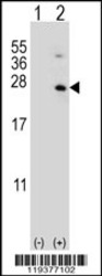
- Western blot analysis of HPRT1 (arrow) using rabbit polyclonal HPRT1 Antibody (C-term) either nontransfected (Lane 1) or lysates transiently transfected transfected (Lane 2) with the HPRT1 gene.
- Sample type
- lysatestransiently transfected with the HPRT1 gene.
- Primary Ab dilution
- 1.0 µg/mL
- Protocol
- Protocol

- Submitted by
- Aviva Systems Biology (provider)
- Main image

- Experimental details
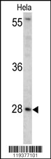
- Western blot analysis of HPRT1 antibody (C-term) in Hela cell lysates (35ug/lane). HPRT1 (arrow) was detected using the purified Pab.
- Sample type
- Hela cell line lysates
- Primary Ab dilution
- 1.0 µg/mL
- Protocol
- Protocol
Supportive validation
- Submitted by
- Aviva Systems Biology (provider)
- Main image
- Experimental details
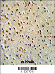
- HPRT1 Antibody (C-term) IHC analysis in formalin fixed and paraffin embedded mouse brain tissue followed by peroxidase conjugation of the secondary antibody and DAB staining. This data demonstrates the use of the HPRT1 Antibody (C-term) for immunohistochemistry. Clinical relevance has not been evaluated.
- Sample type
- mouse brain tissue lysates
- Primary Ab dilution
- 1.0 µg/mL
- Protocol
- Protocol

Supportive validation
- Submitted by
- Aviva Systems Biology (provider)
- Main image
- Experimental details
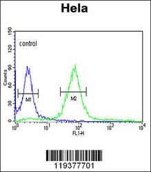
- HPRT1 Antibody (C-term) flow cytometric analysis of Hela cells (right histogram) compared to a negative control cell (left histogram).FITC-conjugated goat-anti-rabbit secondary antibodies were used for the analysis.
- Sample type
- Hela cells
- Primary Ab dilution
- 1.0 µg/mL
- Protocol
- Protocol
