Explore
 Validate
 Learn
Learn Western blot
Antibody data
- Antibody Data
- Antigen structure
- References [2]
- Comments [0]
- Validations
- Western blot [2]
- Immunocytochemistry [1]
- Immunohistochemistry [3]
- Other assay [2]
Submit
Validation data
Reference
Comment
Report error
- Product number
- PA5-88657 - Provider product page

- Provider
- Invitrogen Antibodies
- Product name
- TIRAP Polyclonal Antibody
- Antibody type
- Polyclonal
- Antigen
- Recombinant full-length protein
- Description
- Immunogen sequence: MASSTSLPAP GSRPKKPLGK MADWFRQTLL KKPKKRPNSP ESTSSDASQP TSQDSPLPPS LSSVTSPSLP PTHASDSGSS RWSKDYDVCV CHSEEDLVAA QDLVSYLEGS TASLRCFLQL RDATPGGAIV SELCQALSSS HCRVLLITPG FLQDPWCKYQ MLQALTEAPG AEGCTIPLLS GLSRAAYPPE LRFMYYVDGR GPDGGFRQVK EAVMRYLQTL S; Positive Samples: HeLa, Mouse lung, Rat liver; Cellular Location: Cell membrane, Cytoplasm, Membrane
- Reactivity
- Human, Mouse, Rat
- Host
- Rabbit
- Isotype
- IgG
- Vial size
- 100 µL
- Concentration
- 0.85 mg/mL
- Storage
- -20° C, Avoid Freeze/Thaw Cycles
Submitted references NLRP3 Inflammasome Priming and Activation Are Regulated by a Phosphatidylinositol-Dependent Mechanism.
Molecular Characteristics, Clinical Implication, and Cancer Immunity Interactions of Pyroptosis-Related Genes in Breast Cancer.
Hamilton C, Olona A, Leishman S, MacDonald-Ramsahai K, Cockcroft S, Larrouy-Maumus G, Anand PK
ImmunoHorizons 2022 Aug 29;6(8):642-659
ImmunoHorizons 2022 Aug 29;6(8):642-659
Molecular Characteristics, Clinical Implication, and Cancer Immunity Interactions of Pyroptosis-Related Genes in Breast Cancer.
Xu D, Ji Z, Qiang L
Frontiers in medicine 2021;8:702638
Frontiers in medicine 2021;8:702638
No comments: Submit comment
Supportive validation
- Submitted by
- Invitrogen Antibodies (provider)
- Main image

- Experimental details
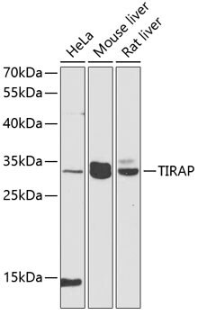
- Western blot analysis of extracts of various cell lines, using TIRAP Polyclonal antibody (Product # PA5-88657) at 1:1000 dilution. Secondary antibody: HRP Goat Anti-Rabbit IgG (H+L) at 1:10000 dilution. Lysates/proteins: 25ug per lane. Blocking buffer: 3% nonfat dry milk in TBST. Exposure time: 90s.

- Submitted by
- Invitrogen Antibodies (provider)
- Main image

- Experimental details
- Western Blot analysis of TIRAP in extracts of various cell lines using TIRAP Polyclonal Antibody (Product # PA5-88657) at a dilution of 1:1000. A HRP Goat Anti-Rabbit IgG (H+L) secondary antibody was used at a dilution of 1:10,000. Lysates/proteins: 25 µg per lane. Blocking buffer: 3% nonfat dry milk in TBST.
Supportive validation
- Submitted by
- Invitrogen Antibodies (provider)
- Main image

- Experimental details
- Immunocytochemistry-Immunofluorescence analysis of TIRAP was performed in U2OS cells using TIRAP Polyclonal Antibody (Product # PA5-88657) at a dilution of 1:100. Blue: DAPI for nuclear staining.

Supportive validation
- Submitted by
- Invitrogen Antibodies (provider)
- Main image

- Experimental details
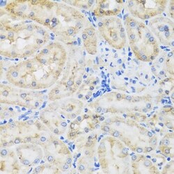
- Immunohistochemistry analysis of TIRAP in paraffin-embedded mouse kidney using TIRAP Polyclonal Antibody (Product # PA5-88657) at a dilution of 1:100.

- Submitted by
- Invitrogen Antibodies (provider)
- Main image
- Experimental details
- Immunohistochemistry analysis of TIRAP in paraffin-embedded rat kidney using TIRAP Polyclonal Antibody (Product # PA5-88657) at a dilution of 1:100.

- Submitted by
- Invitrogen Antibodies (provider)
- Main image

- Experimental details
- Immunohistochemistry analysis of TIRAP in paraffin-embedded mouse liver using TIRAP Polyclonal Antibody (Product # PA5-88657) at a dilution of 1:100.

Supportive validation
- Submitted by
- Invitrogen Antibodies (provider)
- Main image

- Experimental details
- Figure 9 Validation of the expression of GSDMC, IL-18 , and TIRAP in normal breast cells (MCF-10A) and breast cancer cells (MDA-MB-231 and HCC70). (A-D) Western blot detecting the expression of GSDMC, IL-18, and TIRAP in MCF-10A, MDA-MB-231, and HCC70 cells. *** p < 0.001; **** p < 0.0001 . (E-G) Immunofluorescence of the expression of GSDMC, IL-18, and TIRAP in MCF-10A, MDA-MB-231, and HCC70 cells. Scale bar, 5 mum; magnification 200 x.

- Submitted by
- Invitrogen Antibodies (provider)
- Main image

- Experimental details
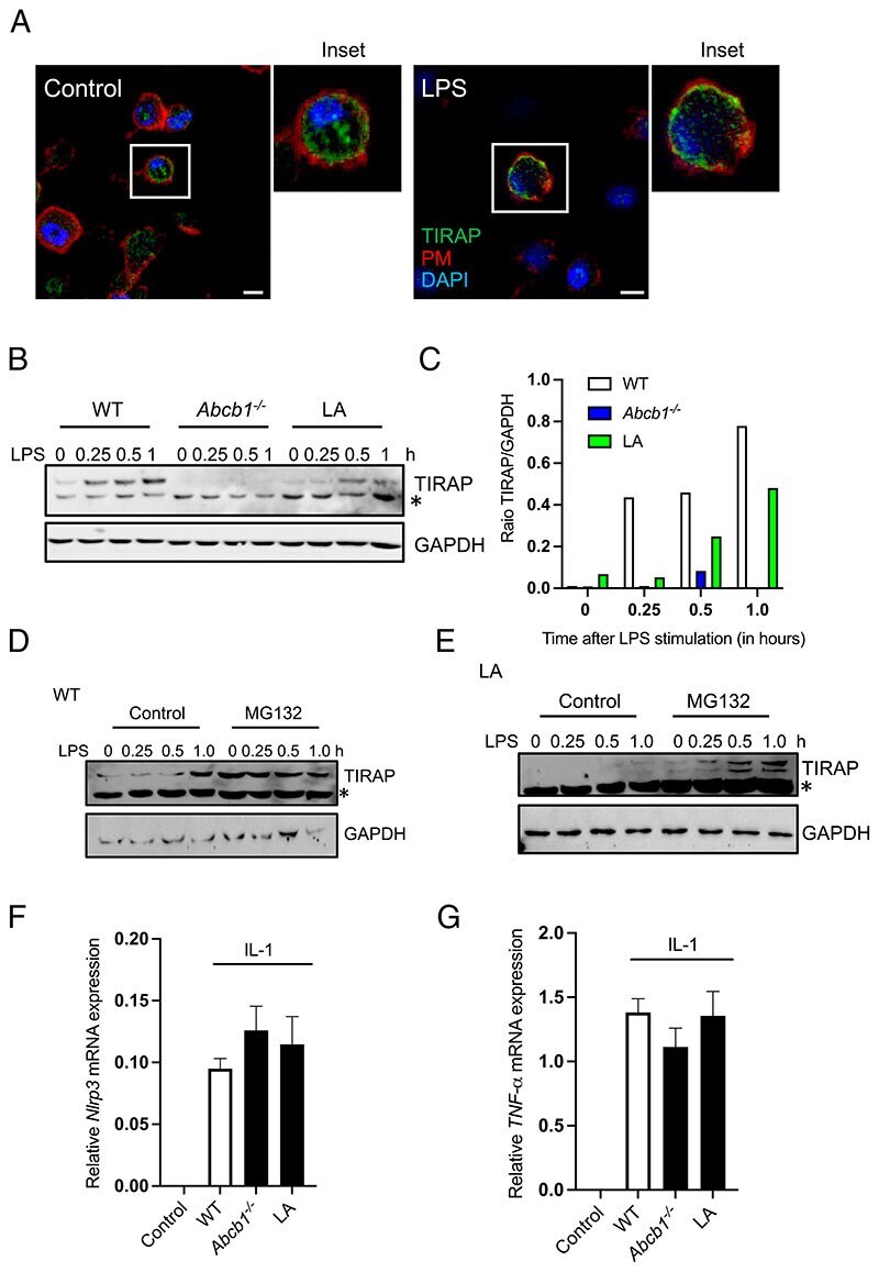
- Figure 6 PI acyl chain profile regulates TIRAP expression. ( A ) Control and LPS-primed WT cells grown on coverslips were labeled with anti-TIRAP Ab, and nuclei were stained with DAPI. The plasma membrane was stained by adding Alexa Fluor 647-conjugated phalloidin for the last 15 min. ( B ) WT, Abcb1 -/- , and LA-supplemented cells were stimulated with LPS for different times. Cell lysates were collected and immunoblotted for TIRAP and GAPDH. ( C ) Quantitation of the WB shown in (B) by ImageJ. ( D and E ) WT (D) and LA-grown (E) cells were pretreated or not with the proteasomal inhibitor MG132 (10 muM) for 30 min before stimulating the cells with LPS for different time points. Cell lysates were collected and immunoblotted for TIRAP and GAPDH. ( F and G ) WT, Abcb1b -/- , and LA-supplemented cells were exposed to IL-1 (2 ng/ml) overnight. RNA was extracted and converted into cDNA. Gene expression of Nlrp3 and TNF-alpha was determined by real-time PCR. The data shown are representative of at least three independent experiments with three to five replicates each. Asterisk (*) on immunoblots denotes a nonspecific band. Scale bars, 5 mum.