 Explore
 Validate
 Learn
Learn Western blot
Antibody data
- Antibody Data
- Antigen structure
- References [0]
- Comments [0]
- Validations
- Western blot [2]
- Immunocytochemistry [1]
- Immunohistochemistry [3]
Submit
Validation data
Reference
Comment
Report error
- Product number
- APR-051-200UL - Provider product page

- Provider
- Invitrogen Antibodies
- Product name
- PTH1R (extracellular) Polyclonal Antibody
- Antibody type
- Polyclonal
- Antigen
- Other
- Description
- Reconstitution: 1 X 50 µL double distilled water (DDW), depending on the sample size. The antibody ships as a lyophilized powder at room temperature. Upon arrival, it should be stored at -20C. The reconstituted solution can be stored at 4C for up to 1 week. For longer periods, small aliquots should be stored at -20C. Avoid multiple freezing and thawing. Centrifuge all antibody preparations before use (10000 x g 5 min).
- Reactivity
- Human, Mouse, Rat
- Host
- Rabbit
- Isotype
- IgG
- Vial size
- 200 µL
- Concentration
- 0.85 mg/mL
- Storage
- -20° C, Avoid Freeze/Thaw Cycles
No comments: Submit comment
Supportive validation
- Submitted by
- Invitrogen Antibodies (provider)
- Main image

- Experimental details
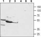
- Western blot analysis of HepG2 (lanes 1 and 4), mouse liver (lanes 2 and 5) and rat liver (lanes 3 and 6) lysates: - 1-3. Anti-PTH1R (extracellular) Antibody (#APR-051), (1:200).4-6. Anti-PTH1R (extracellular) Antibody , preincubated with PTH1R (extracellular) Blocking Peptide (#BLP-PR051).
- Submitted by
- Invitrogen Antibodies (provider)
- Main image

- Experimental details
- Western blot analysis of HepG2 (lanes 1 and 4), mouse liver (lanes 2 and 5) and rat liver (lanes 3 and 6) lysates: - 1-3. Anti-PTH1R (extracellular) Antibody (#APR-051), (1:200).4-6. Anti-PTH1R (extracellular) Antibody , preincubated with PTH1R (extracellular) Blocking Peptide (#BLP-PR051).

Supportive validation
- Submitted by
- Invitrogen Antibodies (provider)
- Main image

- Experimental details
- Expression of Parathyroid hormone receptor 1 in rat U-87 MG cells - Cell surface detection of Parathyroid hormone receptor 1in intact living rat U-87 MG cells. A. Extracellular staining of cells using Anti-PTH1R (extracellular) Antibody (#APR-051), (1:50), (green). B. Merge of A with live view of the cells.

Supportive validation
- Submitted by
- Invitrogen Antibodies (provider)
- Main image

- Experimental details
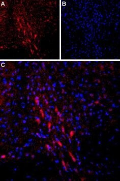
- Expression of Parathyroid hormone receptor 1 in rat brain - Immunohistochemical staining of rat dorsal Raphe nucleus using Anti-PTH1R (extracellular) Antibody (#APR-051). A. PTH1R (red) staining is detected in Raphe neurons (arrows). B. Nucleus staining using DAPI as the counterstain (blue). C. Merged images of A and B.

- Submitted by
- Invitrogen Antibodies (provider)
- Main image

- Experimental details
- Expression of Parathyroid hormone receptor 1 in rat brain - Immunohistochemical staining of rat dorsal Raphe nucleus using Anti-PTH1R (extracellular) Antibody (#APR-051). A. PTH1R (red) staining is detected in Raphe neurons (arrows). B. Nucleus staining using DAPI as the counterstain (blue). C. Merged images of A and B.
- Submitted by
- Invitrogen Antibodies (provider)
- Main image

- Experimental details
- Multiplex staining ofParathyroid hormone receptor 1andOrexin receptor 1in rat ventromedial hypothalamus - Immunohistochemical staining of perfusion-fixed frozen rat brain sections using Anti-Orexin Receptor 1-ATTO Fluor-488 Antibody (#AOR-001-AG), (1:60) and Anti-PTH1R (extracellular) Antibody (#APR-051), (1:60). A. Sections were incubated with Anti-PTH1R (extracellular) Antibody , followed by donkey- Anti-rabbit-Cy3 (red). B. The same sections were incubated with Anti-Orexin Receptor 1-ATTO Fluor-488 Antibody , (green). C. Merge of A and B shows staining of Orexin receptor 1 in nerve fibers (vertical arrows) while Parathyroid hormone receptor 1 is apparent on cell bodies (horizontal arrows). Cell nuclei are stained with DAPI (blue).
