Explore
 Validate
 Learn
Learn Western blot
Antibody data
- Antibody Data
- Antigen structure
- References [0]
- Comments [0]
- Validations
- Western blot [4]
- Immunocytochemistry [1]
- Immunohistochemistry [1]
- Flow cytometry [1]
Submit
Validation data
Reference
Comment
Report error
- Product number
- AP12376PU-N - Provider product page

- Provider
- Acris Antibodies GmbH
- Proper citation
- Acris Antibodies GmbH Cat#AP12376PU-N, RRID:AB_1769179
- Product name
- anti LGR5 / GPR49 (Loop2)
- Antibody type
- Polyclonal
- Antigen
- This antibody is generated from rabbits immunized with a KLH conjugated synthetic peptide between 689-719 amino acids from the loop2 region of Human LGR5/GPR49.
- Reactivity
- Human, Mouse
- Host
- Rabbit
- Vial size
- 0.4 ml
- Concentration
- 0.5 mg/ml
No comments: Submit comment
Supportive validation
- Submitted by
- Acris Antibodies GmbH (provider)
- Main image

- Experimental details
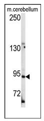
- Western blot analysis of anti-LGR5/GPR49 Antibody (loop2) (Cat.#AP12376PU-N) in mouse cerebellum tissue lysates (35ug/lane).LGR5/GPR49(arrow) was detected using the purified Pab.
- Submitted by
- Acris Antibodies GmbH (provider)
- Main image
- Experimental details
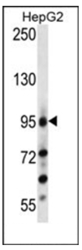
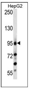
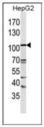
- Western blot analysis of anti-LGR5/GPR49 Antibody (loop2) (Cat.#AP12376PU-N) in in HepG2 cell line lysates (35ug/lane). This demonstrates the LGR5/GPR49 antibody detected the LGR5/GPR49 protein (arrow).
- Submitted by
- Acris Antibodies GmbH (provider)
- Main image
- Experimental details
- Western blot analysis of anti-LGR5/GPR49 Antibody (loop2) (Cat.#AP12376PU-N) in in HepG2 cell line lysates (35ug/lane). This demonstrates the LGR5/GPR49 antibody detected the LGR5/GPR49 protein (arrow).(Kindly offered by Dr. Li).

- Submitted by
- Acris Antibodies GmbH (provider)
- Main image
- Experimental details
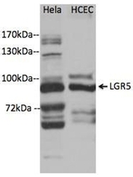
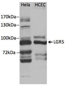
- Western blot analysis of anti-LGR5/GPR49 Antibody (loop2) (Cat.#AP12376PU-N) in Hela and HCEC cell line lysates. This demonstrates the LGR5/GPR49 antibody detected the LGR5/GPR49 protein (arrow) (Kindly offered by Dr. Xiaolin Zhou from UNMC-Eppley Institute).
Supportive validation
- Submitted by
- Acris Antibodies GmbH (provider)
- Main image
- Experimental details
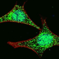
- Fluorescent confocal image of HeLa cells stained with LGR5 (loop2) antibody AP12376PU-N. HeLa cells were fixed with 4% PFA (20 min), permeabilized with Triton X-100 (0.2%, 30 min). Cells were then incubated with AP2745d LGR5 (loop2) primary antibody (1:100, 2 h at room temperature). For secondary antibody, Alexa Fluor® 488 conjugated donkey anti-rabbit antibody (green) was used (1:1000, 1h). Nuclei were counterstained with Hoechst 33342 (blue) (10 ?g/ml, 5 min). Note the highly specific localization of the LGR5 immunosignal to the nucleus and cytoplasm, supported by Human Protein Atlas Data

Supportive validation
- Submitted by
- Acris Antibodies GmbH (provider)
- Main image
- Experimental details
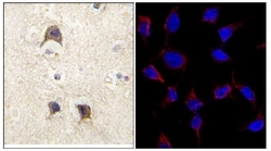
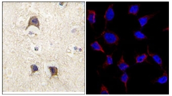
- (LEFT) Formalin-fixed and paraffin-embedded human brain tissue reacted with LGR5/GPR49 antibody (loop2) (Cat.#AP12376PU-N), which was peroxidase-conjugated to the secondary antibody, followed by DAB staining. This data demonstrates the use of this antibody for Immunohistochemistry. Clinical relevance has not been evaluated. (RIGHT) Immunofluorescence analysis of anti-LGR5/GPR49 Antibody (loop2) (Cat#AP12376PU-N) in HeLa cells. 0.025 mg/ml primary antibody was followed by Alexa-Fluor-546-conjugated Donkey anti-Rabbit lgG (H+L). Alexa-Fluor-546 emits orange Fluorescence.
Supportive validation
- Submitted by
- Acris Antibodies GmbH (provider)
- Main image

- Experimental details
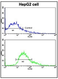
- Flow Cytometric analysis of HepG2 cells using LGR5/GPR49 Antibody (loop2) (bottom histogram) compared to a negative control cell (top histogram). FITC-conjugated Goat-anti-Rabbit secondary antibodies were used for the analysis.