 Explore
 Validate
 Learn
Learn Western blot
Antibody data
- Antibody Data
- Antigen structure
- References [0]
- Comments [0]
- Validations
- Western blot [1]
- Immunocytochemistry [3]
- Immunohistochemistry [2]
Submit
Validation data
Reference
Comment
Report error
- Product number
- ACC-049-25UL - Provider product page

- Provider
- Invitrogen Antibodies
- Product name
- TRPM8 (extracellular) Polyclonal Antibody
- Antibody type
- Polyclonal
- Antigen
- Other
- Description
- Reconstitution: 1 X 25 µL double distilled water (DDW), depending on the sample size. The antibody ships as a lyophilized powder at room temperature. Upon arrival, it should be stored at -20C. The reconstituted solution can be stored at 4C for up to 1 week. For longer periods, small aliquots should be stored at -20C. Avoid multiple freezing and thawing. Centrifuge all antibody preparations before use (10000 x g 5 min).
- Reactivity
- Human, Mouse, Rat
- Host
- Rabbit
- Isotype
- IgG
- Vial size
- 25 µL
- Concentration
- 0.8 mg/mL
- Storage
- -20° C, Avoid Freeze/Thaw Cycles
No comments: Submit comment
Supportive validation
- Submitted by
- Invitrogen Antibodies (provider)
- Main image

- Experimental details
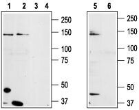
- Western blot analysis of prostate carcinoma cell lines DU145 (lanes 1 and 3),Human LNCaP prostate carcinoma (lanes 2 and 4) and mouse-TRPM8 transfected HEK-293 (lanes 5 and 6) cell lysates: - 1,2,5. Anti-TRPM8 (extracellular) Antibody (#ACC-049), (1:200).3,4,6. Anti-TRPM8 (extracellular) Antibody , preincubated with TRPM8 (extracellular) Blocking Peptide (#BLP-CC049).
Supportive validation
- Submitted by
- Invitrogen Antibodies (provider)
- Main image

- Experimental details
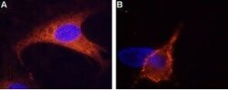
- Expression of TRPM8 in rat DRG cells - Immunocytochemistry of rat dorsal root ganglion (DRG) cells. A. Intracellular staining of cells with Anti-TRPM8 (extracellular) Antibody (#ACC-049), (1:500) followed by goat Anti-rabbit-AlexaFluor-555 secondary Antibody . B. Extracellular staining of live cells with Anti-TRPM8 (extracellular) Antibody (1:50) followed by goat Anti-rabbit-AlexaFluor-555 secondary Antibody . The cell-permeable dye Hoechst 33342 (blue) was used for nuclear staining.

- Submitted by
- Invitrogen Antibodies (provider)
- Main image
- Experimental details
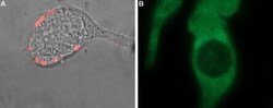
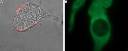
- Expression of TRPM8 in LNCaP prostate carcinoma cell line - Cell surface detection of TRPM8 in LNCaP cells with Anti-TRPM8 (extracellular) Antibody (#ACC-049). A. Extracellular staining of intact LNCaP cells with Anti-TRPM8 (extracellular) Antibody (1:100), followed by goat Anti-rabbit-AlexaFluor-550 (red), (x100). B. Intracellular staining of LNCaP cells with Anti-TRPM8 (extracellular) Antibody (1:1000), followed by goat Anti-rabbit-AlexaFluor-488 (green), (x100).
Supportive validation
- Submitted by
- Invitrogen Antibodies (provider)
- Main image
- Experimental details
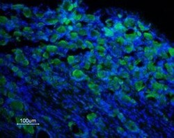
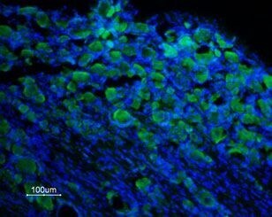
- Expression of TRPM8 in rat DRG - Immunohistochemical staining of rat dorsal root ganglion (DRG) frozen sections using Anti-TRPM8 (extracellular) Antibody (#ACC-049), (1:100). TRPM8 is expressed in DRG neurons. Hoechst 33342 is used as the counterstain.
- Submitted by
- Invitrogen Antibodies (provider)
- Main image
- Experimental details
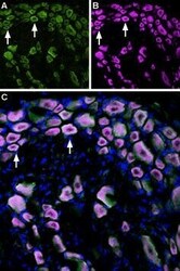
- Multiplex staining of TRPM8 and TrkA in rat DRG - Immunohistochemical staining of perfusion-fixed frozen rat dorsal root ganglia (DRG) sections using Anti-TrkA (extracellular)-ATTO Fluor-633 Antibody (#ANT-018-FR), (1:60) and Anti-TRPM8 (extracellular) Antibody (#ACC-049), (1:300). A. TRPM8 labeling followed by goat- Anti-rabbit-Alexa-488 (green). B. The same section was then labeled for TrkA (purple). C. Merge of A and B demonstrates co-localization of TRPM8 and TrkA in rat DRG (arrows). Cell nuclei were stained with DAPI (blue).
