 Explore
 Validate
 Learn
Learn Western blot
Antibody data
- Antibody Data
- Antigen structure
- References [1]
- Comments [0]
- Validations
- Western blot [1]
- Immunocytochemistry [1]
- Immunoprecipitation [1]
- Immunohistochemistry [9]
Submit
Validation data
Reference
Comment
Report error
- Product number
- GTX83458 - Provider product page

- Provider
- GeneTex
- Proper citation
- GeneTex Cat#GTX83458, RRID:AB_10729059
- Product name
- TYRO3 antibody [2C4]
- Antibody type
- Monoclonal
- Reactivity
- Human
- Host
- Mouse
Submitted references Ampelopsins A and C Induce Apoptosis and Metastasis through Downregulating AxL, TYRO3, and FYN Expressions in MDA-MB-231 Breast Cancer Cells.
Huang C, Huang YL, Wang CC, Pan YL, Lai YH, Huang HC
Journal of agricultural and food chemistry 2019 Mar 13;67(10):2818-2830
Journal of agricultural and food chemistry 2019 Mar 13;67(10):2818-2830
No comments: Submit comment
Supportive validation
- Submitted by
- GeneTex (provider)
- Main image

- Experimental details
- HEK293T cells were transfected with the pCMV6-ENTRY control (Left lane) or pCMV6-ENTRY TYRO3 (Right lane) cDNA for 48 hrs and lysed. Equivalent amounts of cell lysates (5 ug per lane) were separated by SDS-PAGE and immunoblotted with anti-TYRO3.

Supportive validation
- Submitted by
- GeneTex (provider)
- Main image

- Experimental details
- Anti-TYRO3 mouse monoclonal antibody (GTX83458) immunofluorescent staining of COS7 cells transiently transfected with TYRO3

Supportive validation
- Submitted by
- GeneTex (provider)
- Main image

- Experimental details
- Immunoprecipitation(IP) of TYRO3 by using TrueMab monoclonal anti-TYRO3 antibodies (Negative control: IP without adding anti-TYRO3 antibody.). For each experiment, 500ul of DDK tagged TYRO3 overexpression lysates (at 1:5 dilution with HEK293T lysate), 2ug of anti-TYRO3 antibody and 20ul (0.1mg) of goat anti-mouse conjugated magnetic beads were mixed and incubated overnight. After extensive wash to remove any non-specific binding, the immuno-precipitated products were analyzed with rabbit anti-DDK polyclonal antibody.

Supportive validation
- Submitted by
- GeneTex (provider)
- Main image

- Experimental details
- Immunohistochemical staining of paraffin-embedded Adenocarcinoma of breast tissue using anti-TYRO3 mouse monoclonal antibody. (GTX83458, Dilution 1:50)

- Submitted by
- GeneTex (provider)
- Main image
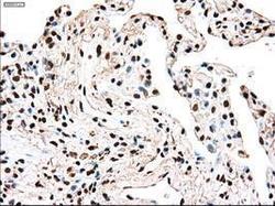
- Experimental details
- Immunohistochemical staining of paraffin-embedded Carcinoma of lung tissue using anti-TYRO3mouse monoclonal antibody. (GTX83458, Dilution 1:50)
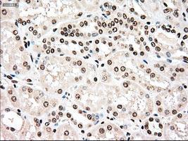
- Submitted by
- GeneTex (provider)
- Main image

- Experimental details
- Immunohistochemical staining of paraffin-embedded Kidney tissue using anti-TYRO3mouse monoclonal antibody. (GTX83458, Dilution 1:50)
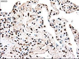
- Submitted by
- GeneTex (provider)
- Main image

- Experimental details
- Immunohistochemical staining of paraffin-embedded lung tissue using anti-TYRO3mouse monoclonal antibody. (GTX83458, Dilution 1:50)

- Submitted by
- GeneTex (provider)
- Main image

- Experimental details
- Immunohistochemical staining of paraffin-embedded Adenocarcinoma of ovary tissue using anti-TYRO3mouse monoclonal antibody. (GTX83458, Dilution 1:50)

- Submitted by
- GeneTex (provider)
- Main image

- Experimental details
- Immunohistochemical staining of paraffin-embedded breast tissue using anti-TYRO3 mouse monoclonal antibody. (GTX83458, Dilution 1:50)

- Submitted by
- GeneTex (provider)
- Main image

- Experimental details
- Immunohistochemical staining of paraffin-embedded Carcinoma of kidney tissue using anti-TYRO3mouse monoclonal antibody. (GTX83458, Dilution 1:50)

- Submitted by
- GeneTex (provider)
- Main image

- Experimental details
- Immunohistochemical staining of paraffin-embedded Ovary tissue using anti-TYRO3mouse monoclonal antibody. (GTX83458, Dilution 1:50)

- Submitted by
- GeneTex (provider)
- Main image

- Experimental details
- Immunohistochemical staining of paraffin-embedded pancreas tissue using anti-TYRO3mouse monoclonal antibody. (GTX83458, Dilution 1:50)
