 Explore
 Validate
 Learn
Learn Western blot
Antibody data
- Antibody Data
- Antigen structure
- References [0]
- Comments [0]
- Validations
- Western blot [2]
- Immunocytochemistry [2]
Submit
Validation data
Reference
Comment
Report error
- Product number
- APC-104-200UL - Provider product page

- Provider
- Invitrogen Antibodies
- Product name
- KCNH1 (EAG-1) Polyclonal Antibody
- Antibody type
- Polyclonal
- Antigen
- Other
- Description
- Reconstitution: 1 X 25 µL double distilled water (DDW), depending on the sample size. The antibody ships as a lyophilized powder at room temperature. Upon arrival, it should be stored at -20C. The reconstituted solution can be stored at 4C for up to 1 week. For longer periods, small aliquots should be stored at -20C. Avoid multiple freezing and thawing. Centrifuge all antibody preparations before use (10000 x g 5 min).
- Reactivity
- Human, Mouse, Rat
- Host
- Rabbit
- Isotype
- IgG
- Vial size
- 200 µL
- Concentration
- 0.6 mg/mL
- Storage
- -20° C, Avoid Freeze/Thaw Cycles
No comments: Submit comment
Supportive validation
- Submitted by
- Invitrogen Antibodies (provider)
- Main image

- Experimental details
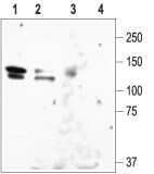
- Western blot analysisof rat brain lysate (lanes 1 and 3) and HEK-KCNH1 (lanes 2 and 4): - 1,2. Anti-KCNH1 (EAG-1) Antibody (#APC-104), (1:200).3,4. Anti-KCNH1 (EAG-1) Antibody , preincubated with KCNH1/EAG-1 Blocking Peptide (#BLP-PC104).
- Submitted by
- Invitrogen Antibodies (provider)
- Main image

- Experimental details
- Western blot analysisof rat brain lysate (lanes 1 and 3) and HEK-KCNH1 (lanes 2 and 4): - 1,2. Anti-KCNH1 (EAG-1) Antibody (#APC-104), (1:200).3,4. Anti-KCNH1 (EAG-1) Antibody , preincubated with KCNH1/EAG-1 Blocking Peptide (#BLP-PC104).

Supportive validation
- Submitted by
- Invitrogen Antibodies (provider)
- Main image

- Experimental details
- Expression of EAG1in human MDA-468 mammary gland adenocarcinoma cells. Immunocytochemical staining of human MDA-468 mammary gland adenocarcinoma cells. A. Cells were stained with Anti-KCNH1 (EAG-1) Antibody (#APC-104), (1:200) followed by goat Anti-rabbit-AlexaFluor-555 secondary Antibody (red). B.Nuclei were visualized with the cell-permeable dye Hoechst 33342 (blue). C.Merged image of panels A and B.
