 Explore
 Validate
 Learn
Learn Western blot
Antibody data
- Antibody Data
- Antigen structure
- References [1]
- Comments [0]
- Validations
- Western blot [3]
- Immunohistochemistry [1]
- Other assay [1]
Submit
Validation data
Reference
Comment
Report error
- Product number
- PA5-36062 - Provider product page

- Provider
- Invitrogen Antibodies
- Product name
- LIMK1/LIMK2 Polyclonal Antibody
- Antibody type
- Polyclonal
- Antigen
- Synthetic peptide
- Description
- This antibody detects endogenous protein at a molecular weight of 72 kDa.
- Concentration
- 1 mg/mL
Submitted references IL-4 Causes Hyperpermeability of Vascular Endothelial Cells through Wnt5A Signaling.
Skaria T, Burgener J, Bachli E, Schoedon G
PloS one 2016;11(5):e0156002
PloS one 2016;11(5):e0156002
No comments: Submit comment
Supportive validation
- Submitted by
- Invitrogen Antibodies (provider)
- Main image

- Experimental details
- Western blot analysis of LIMK1/LIMK2 in Lane 1: HEK293T whole cell lysate (40 µg), Lane 2: Panc1 whole cell lysate (40 µg), Lane 3: A549 whole cell lysate (40 µg), Lane 4: PC12 whole cell lysate (40 µg), Lane 5: AML-12 whole cell lysate (40 µg). Samples were incubated with LIMK1/LIMK2 polyclonal antibody (Product # PA5-36062) at a dilution of 1:500.

- Submitted by
- Invitrogen Antibodies (provider)
- Main image

- Experimental details
- Western blot analysis of LIMK1+LIMK2 using LIMK1+LIMK2 polyclonal antibody (Product # PA5-36062) at a dilution of 1:500. Lane 1: HEK293T whole cell lysate, Lane 2: sp2/0 whole cell lysate, Lane 3: H9C2 whole cell lysate.

Supportive validation
- Submitted by
- Invitrogen Antibodies (provider)
- Main image
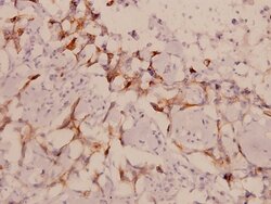
- Experimental details
- Immunohistochemistry analysis of LIMK1/LIMK2 in paraffin-embedded human breast carcinoma tissue. Samples were incubated with LIMK1/LIMK2 polyclonal antibody (Product # PA5-36062) at a dilution of 1:100.

Supportive validation
- Submitted by
- Invitrogen Antibodies (provider)
- Main image

- Experimental details
- Fig 4 Phosphorylation of LIMK2 and CFL1 in IL-4 treated HCAEC. (A) pLIMK2 (pT505) and pCFL1 (pCofilin-1, pS3) were immunostained (red) in HCAEC treated with IL-4 either alone or in combination with Y-27632. F-actin (fluorescent phalloidin, green) and nuclei (DAPI, blue) were also stained. Photomicrographs were acquired using a Zeiss Axioskope equipped with AxioCam MRm digital camera and AxioVision Rel.4.6 software. Original magnification, 200x. (B) Mean cellular fluorescent intensities of pLIMK2 and pCFL1. Data represent mean +- SD from five different regions. * P
