Explore
 Validate
 Learn
Learn Western blot
Antibody data
- Antibody Data
- Antigen structure
- References [1]
- Comments [0]
- Validations
- Western blot [1]
- Immunohistochemistry [1]
- Other assay [1]
Submit
Validation data
Reference
Comment
Report error
- Product number
- PA5-19228 - Provider product page

- Provider
- Invitrogen Antibodies
- Product name
- Defensin 1/3 Polyclonal Antibody
- Antibody type
- Polyclonal
- Antigen
- Synthetic peptide
- Reactivity
- Human
- Host
- Goat
- Isotype
- IgG
- Vial size
- 100 µg
- Concentration
- 0.5 mg/mL
- Storage
- -20° C, Avoid Freeze/Thaw Cycles
Submitted references Quantitative proteomic characterization of microvesicles/exosomes from the cerebrospinal fluid of patients with acute bilirubin encephalopathy.
Tan N, Hu S, Hu Z, Wu Z, Wang B
Molecular medicine reports 2020 Aug;22(2):1257-1268
Molecular medicine reports 2020 Aug;22(2):1257-1268
No comments: Submit comment
Supportive validation
- Submitted by
- Invitrogen Antibodies (provider)
- Main image

- Experimental details
- Western Blot staining of Human Spleen lysate using Product # PA5-19228 at a concentration of 2 µg/mL, the primary antibody incubation was 1 hour and the detection method was chemiluminescence.

Supportive validation
- Submitted by
- Invitrogen Antibodies (provider)
- Main image

- Experimental details
- Immunohistochemical analysis of Defensin 1 3 in paraffin embedded human thymus using a Defensin 1 3 polyclonal antibody (Product #PA5-19228) at a concentration of 3.8 µg/mL. Steamed antigen retrieval was performed with pH 6 buffered citrate. Samples were then stained with alkaline phosphatase.

Supportive validation
- Submitted by
- Invitrogen Antibodies (provider)
- Main image

- Experimental details
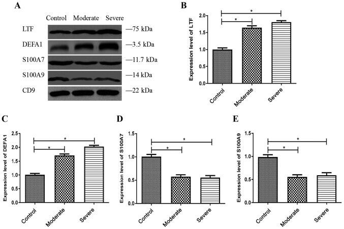
- Figure 6. Verification of differentially expressed proteins using western blotting. (A) Representative western blots demonstrate the expression levels of LTF, DEFA1, S100A7 and S100A9 in the MV/E isolated from the CSF. Histogram data shows the densitometric semi-quantification (mean +- SD) of (B) LTF, (C) DEFA1, (D) S100A7 and (E) S100A9 expression levels. Protein densitometric semi-quantification was normalized to the density of CD9 protein. CD9 is an exosome surface marker. *P