 Explore
 Validate
 Learn
Learn Western blot
Antibody data
- Antibody Data
- Antigen structure
- References [1]
- Comments [0]
- Validations
- Western blot [2]
- Immunohistochemistry [1]
- Other assay [1]
Submit
Validation data
Reference
Comment
Report error
- Product number
- PA5-47890 - Provider product page

- Provider
- Invitrogen Antibodies
- Product name
- EBF2 Polyclonal Antibody
- Antibody type
- Polyclonal
- Antigen
- Recombinant full-length protein
- Description
- In direct ELISAs, less than 1% cross-reactivity with recombinant mouse (rm) EBF-1 and rmEBF-3 is observed.
- Concentration
- 0.2 mg/mL
Submitted references Spinal gastrin releasing peptide receptor expressing interneurons are controlled by local phasic and tonic inhibition.
Freitag FB, Ahemaiti A, Jakobsson JET, Weman HM, Lagerström MC
Scientific reports 2019 Nov 12;9(1):16573
Scientific reports 2019 Nov 12;9(1):16573
No comments: Submit comment
Supportive validation
- Submitted by
- Invitrogen Antibodies (provider)
- Main image

- Experimental details
- Western blot analysis from lysates of 3T3-L1 mouse embryonic fibroblast adipose-like cell line and HepG2 human hepatocellular carcinoma cell line. PVDF membrane was probed with 1 µg/mL of Sheep Anti-human/mouse EBF-2 Antigen Affinity-purified Polyclonal Antibody (Product # PA5-47890) followed by HRP-conjugated Anti-Sheep IgG Secondary Antibody. A specific band was detected for EBF-2 at approximately 65 kDa (as indicated). This experiment was conducted under reducing conditions.

- Submitted by
- Invitrogen Antibodies (provider)
- Main image

- Experimental details
- Western blot analysis of EBF2 in 3T3‚L1 mouse embryonic fibroblast adipose-like cell line and HepG2 human hepatocellular carcinoma cell line. Samples were incubated in EBF2 polyclonal antibody (Product # PA5-47890) using a dilution of 1 µg/mL followed by a HRP-conjugated Anti-Sheep IgG secondary antibody. A specific band was detected for EBF-2 at approximately 65 kDa (as indicated). This experiment was conducted under reducing conditions.

Supportive validation
- Submitted by
- Invitrogen Antibodies (provider)
- Main image

- Experimental details
- Immunohistochemical analysis of EBF2 in immersion fixed frozen sections of mouse embryo (15 d.p.c.). Samples were incubated in EBF2 polyclonal antibody (Product # PA5-47890) using a dilution of 10 µg/mL overnight at 4 °C. Tissue was stained using the Anti-Sheep HRP-DAB Cell & Tissue Staining Kit (brown) and counterstained with hematoxylin (blue). Specific staining was localized to developing muscle cells.

Supportive validation
- Submitted by
- Invitrogen Antibodies (provider)
- Main image
- Experimental details
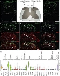
- Figure 1 The Grpr-Cre population of spinal interneurons is predominantly excitatory. Analysis of adult Grpr-Cre neurons displayed through a viral reporter system. ( a ) Coronal section of lumbar spinal cord annotated using Rexed laminae following an injection of AAV2-EF1a-DIO-EYFP virus using the angled injection scheme. Grpr-Cre neurons in green. ( b ) Illustrating the difference between the angled and the straight injection schemes. ( c ) Coronal section of lumbar spinal cord following an injection of the reporter virus AAVDJ-EF1a-DIO-HTB using the straight injection scheme. Grpr-Cre neurons in green. ( d-f ) Immunohistochemistry against PAX2 (d), TLX3 (e) and EBF2 (f) with Grpr-Cre neurons in green and the antigen in red. Dashed lines outline lamina I-IV. Arrows indicate Grpr-Cre neurons overlapping with the antigen. ( g ) Grpr neurons express both excitatory and inhibitory markers and genes important for glutamatergic, GABAergic and glycinergic signaling input. Violin plot of normalized expression of marker genes and AMPA, GABA receptor type A and glycine receptor subunits in Grpr -expressing neurons. For gene expression of all presented genes in all Haring cell types see Figure S3A . For gene expression in all Grpr neurons grouped by Haring cell types, see Figure S3B . Scale bars in ( a ) and ( c ) correspond to 200 um while scale bars in ( d-f ) correspond to 100 um.
