Explore
 Validate
 Learn
Learn Western blot
Antibody data
- Antibody Data
- Antigen structure
- References [5]
- Comments [0]
- Validations
- Western blot [1]
- Immunohistochemistry [1]
Submit
Validation data
Reference
Comment
Report error
- Product number
- AF7006 - Provider product page

- Provider
- R&D Systems
- Product name
- Human/Mouse EBF-2 Antibody
- Antibody type
- Polyclonal
- Description
- Immunogen affinity purified. Detects mouse EBF-2 in direct ELISAs. Detects mouse and human EBF-2 in Western blots. In direct ELISAs, less than 1% cross-reactivity with recombinant mouse (rm) EBF-1 and rmEBF-3 is observed.
- Reactivity
- Human, Mouse
- Host
- Sheep
- Conjugate
- Unconjugated
- Antigen sequence
O08792- Isotype
- IgG
- Vial size
- 100 ug
- Concentration
- LYOPH
- Storage
- Use a manual defrost freezer and avoid repeated freeze-thaw cycles. 12 months from date of receipt, -20 to -70 °C as supplied. 1 month, 2 to 8 °C under sterile conditions after reconstitution. 6 months, -20 to -70 °C under sterile conditions after reconstitution.
Submitted references The tumor secretory factor ZAG promotes white adipose tissue browning and energy wasting.
A Renewable Source of Human Beige Adipocytes for Development of Therapies to Treat Metabolic Syndrome.
Id1 Promotes Obesity by Suppressing Brown Adipose Thermogenesis and White Adipose Browning.
A Multi-step Transcriptional and Chromatin State Cascade Underlies Motor Neuron Programming from Embryonic Stem Cells.
A long noncoding RNA transcriptional regulatory circuit drives thermogenic adipocyte differentiation.
Elattar S, Dimri M, Satyanarayana A
FASEB journal : official publication of the Federation of American Societies for Experimental Biology 2018 Sep;32(9):4727-4743
FASEB journal : official publication of the Federation of American Societies for Experimental Biology 2018 Sep;32(9):4727-4743
A Renewable Source of Human Beige Adipocytes for Development of Therapies to Treat Metabolic Syndrome.
Su S, Guntur AR, Nguyen DC, Fakory SS, Doucette CC, Leech C, Lotana H, Kelley M, Kohli J, Martino J, Sims-Lucas S, Liaw L, Vary C, Rosen CJ, Brown AC
Cell reports 2018 Dec 11;25(11):3215-3228.e9
Cell reports 2018 Dec 11;25(11):3215-3228.e9
Id1 Promotes Obesity by Suppressing Brown Adipose Thermogenesis and White Adipose Browning.
Patil M, Sharma BK, Elattar S, Chang J, Kapil S, Yuan J, Satyanarayana A
Diabetes 2017 Jun;66(6):1611-1625
Diabetes 2017 Jun;66(6):1611-1625
A Multi-step Transcriptional and Chromatin State Cascade Underlies Motor Neuron Programming from Embryonic Stem Cells.
Velasco S, Ibrahim MM, Kakumanu A, Garipler G, Aydin B, Al-Sayegh MA, Hirsekorn A, Abdul-Rahman F, Satija R, Ohler U, Mahony S, Mazzoni EO
Cell stem cell 2017 Feb 2;20(2):205-217.e8
Cell stem cell 2017 Feb 2;20(2):205-217.e8
A long noncoding RNA transcriptional regulatory circuit drives thermogenic adipocyte differentiation.
Zhao XY, Li S, Wang GX, Yu Q, Lin JD
Molecular cell 2014 Aug 7;55(3):372-82
Molecular cell 2014 Aug 7;55(3):372-82
No comments: Submit comment
Supportive validation
- Submitted by
- R&D Systems (provider)
- Main image

- Experimental details
- Detection of Human and Mouse EBF-2 by Western Blot. Western blot shows lysates of 3T3-L1 mouse embryonic fibroblast adipose-like cell line and HepG2 human hepatocellular carcinoma cell line. PVDF membrane was probed with 1 µg/mL of Sheep Anti-Human/Mouse EBF-2 Antigen Affinity-purified Polyclonal Antibody (Catalog # AF7006) followed by HRP-conjugated Anti-Sheep IgG Secondary Antibody (Catalog # HAF016). A specific band was detected for EBF-2 at approximately 65 kDa (as indicated). This experiment was conducted under reducing conditions and using Immunoblot Buffer Group 8.

Supportive validation
- Submitted by
- R&D Systems (provider)
- Main image

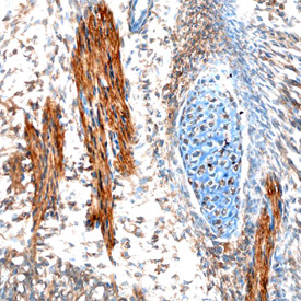
- Experimental details
- EBF-2 in Mouse Embryo. EBF-2 was detected in immersion fixed frozen sections of mouse embryo (15 d.p.c.) using Sheep Anti-Mouse EBF-2 Antigen Affinity-purified Polyclonal Antibody (Catalog # AF7006) at 10 µg/mL overnight at 4 °C. Tissue was stained using the Anti-Sheep HRP-DAB Cell & Tissue Staining Kit (brown; Catalog # CTS019) and counterstained with hematoxylin (blue). Specific staining was localized to developing muscle cells. View our protocol for Chromogenic IHC Staining of Frozen Tissue Sections.