 Explore
 Validate
 Learn
Learn Western blot
Antibody data
- Antibody Data
- Antigen structure
- References [0]
- Comments [0]
- Validations
- Western blot [2]
- Immunocytochemistry [4]
- Immunohistochemistry [6]
Submit
Validation data
Reference
Comment
Report error
- Product number
- GTX37364 - Provider product page

- Provider
- GeneTex
- Proper citation
- GeneTex Cat#GTX37364, RRID:AB_11168027
- Product name
- CD303 antibody
- Antibody type
- Polyclonal
- Reactivity
- Mouse, Rat, Porcine
- Host
- Rabbit
No comments: Submit comment
Supportive validation
- Submitted by
- GeneTex (provider)
- Main image

- Experimental details
- Western blot analysis of Rat small intestine tissue using CD303 antibody (dilution at 2 ug/mL)

- Submitted by
- GeneTex (provider)
- Main image

- Experimental details
- WB analysis of 293T cells (lane 1), Hela cells (lane 2), human ovarian cancer (lane 3), human thyroid cancer (lane 4) using CD303 antibody (I ug/ml)

Supportive validation
- Submitted by
- GeneTex (provider)
- Main image

- Experimental details
- IF analysis of mouse lymph gland tissue using CD303 antibody (dilution of primary antibody at 1:100)

- Submitted by
- GeneTex (provider)
- Main image

- Experimental details
- Immunofluorescence analysis of mouse lymph gland tissue using anti-CD303 (dilution of primary antibody - 1:100)

- Submitted by
- GeneTex (provider)
- Main image

- Experimental details
- IF image of mouse lymph gland tissue using CD303 antibody (primary antibody at 1:100)

- Submitted by
- GeneTex (provider)
- Main image

- Experimental details
- Immunofluorescence image of mouse lymph gland tissue using anti-CD303 (dilution at 1:100)

Supportive validation
- Submitted by
- GeneTex (provider)
- Main image
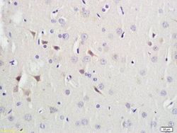
- Experimental details
- IHC-P analysis of mouse brain tissue using CD303 antibody.

- Submitted by
- GeneTex (provider)
- Main image

- Experimental details
- IHC-P image of mouse lymph gland tissue using anti-CD303 (dilution of primary antibody at 1:100)

- Submitted by
- GeneTex (provider)
- Main image

- Experimental details
- Immunohistochemical staining of paraffin embedded mouse lymph gland tissue using anti-CD303 (primary antibody at 1:200)

- Submitted by
- GeneTex (provider)
- Main image
- Experimental details
- IHC-P image of mouse lymph gland tissue using CD303 antibody (dilution of primary antibody at 1:200)

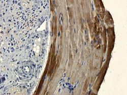
- Submitted by
- GeneTex (provider)
- Main image

- Experimental details
- Immunohistochemical analysis of paraffin-embedded pig small intestine vessel tissue using CD303 antibody (dilution at 1:100)

- Submitted by
- GeneTex (provider)
- Main image

- Experimental details
- IHC-P staining of pig small intestine tissue using anti-CD303 (dilution at 1:100)
