Explore
 Validate
 Learn
Learn Western blot
Antibody data
- Antibody Data
- Antigen structure
- References [0]
- Comments [0]
- Validations
- Western blot [1]
- Flow cytometry [4]
Submit
Validation data
Reference
Comment
Report error
- Product number
- NBP2-26649 - Provider product page

- Provider
- Novus Biologicals
- Product name
- Mouse Monoclonal KLRC1 Antibody
- Antibody type
- Monoclonal
- Antigen
- Animals were immunized with a synthetic peptide from the alpha-2 domain of Qa-1b. Qa-1 is a Class Ib molecule recognized by cytotoxic T cells and natural killer cells. Spleen cells were fused with P3X63Ag8.653 myeloma cells.The antibody reacts with the Qa-1b Class Ib molecule.
- Reactivity
- Mouse
- Host
- Mouse
- Isotype
- IgG
- Vial size
- 0.1mg
- Storage
- Store at 4°C. Do not freeze.
No comments: Submit comment
Supportive validation
- Submitted by
- Novus Biologicals (provider)
- Main image
- Experimental details
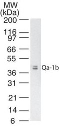
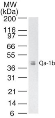
- Western Blot: QA1b Antibody (6A8.6F10.1A6) [NBP2-26649] - Analysis of QA1b in mouse thymus tissue lysate using NBP2-26649 at 2 ug/mL.
Supportive validation
- Submitted by
- Novus Biologicals (provider)
- Main image
- Experimental details
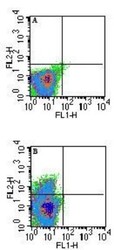
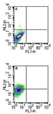
- Flow Cytometry: QA1b Antibody (6A8.6F10.1A6) [NBP2-26649] - Analysis of QA1b in unstimulated Balb/c mouse splenocytes using A) isotype control and B) NBP2-26649 (with an anti-mouse IgG1-PE secondary) at 0.25 ug/10^6 cells. A shift is seen in only a small percentage of cells.

- Submitted by
- Novus Biologicals (provider)
- Main image
- Experimental details
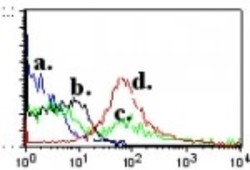
- Flow Cytometry: QA1b Antibody (6A8.6F10.1A6) [NBP2-26649] - Analysis using the Biotin conjugate of NBP2-26649. Staining of resting and ConA activated (5 ug/mL for 72 hr) BALB/C splenocytes using 0.5 ug of Qa-1b biotinylated antibody. CD3 positive cells were gated. Streptavidin - PE (SA-PE) was used as secondary antibody. a) SA-PE resting cells. b) SA-PE ConA activated cells. c) Qa-1b resting cells. d) Qa-1b ConA activated cells.

- Submitted by
- Novus Biologicals (provider)
- Main image

- Experimental details
- Flow Cytometry: QA1b Antibody (6A8.6F10.1A6) [NBP2-26649] - Analysis of Qa-1b in 72 hour ConA-stimulated Balb/c mouse splenocytes using A) isotype control and B) NBP2-26649 (with an anti-mouse IgG1-PE secondary) at 0.25 ug/10^6 cells. A shift is seen in the entire population of cells Image using the Azide Free form of this antibody.
- Submitted by
- Novus Biologicals (provider)
- Main image
- Experimental details
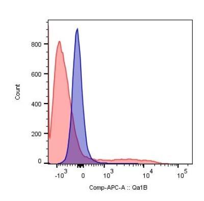
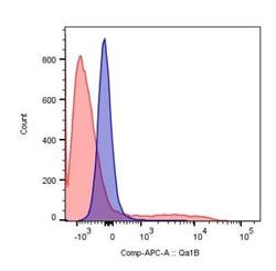
- Flow Cytometry: QA1b Antibody (6A8.6F10.1A6) [NBP2-26649] - Experimental autoimmune encephalomyelitis was induced in C57BL6/J mice, and cells from central nervous system (brain+spinal cord) were isolated when the animals were at the peak of the disease. Cells were stained for QA1b, CD8, CD4, TNF, IFNg, IL-17, plus for viability to exclude dead cells. CNS cells of EAE mice stained for viable cells using APC conjugated QA1B antibody (NBP2-26649APC). Viable, CD8+ cells were gated and QA1b staining is showed in control cells, non-stained for QA1b (blue), and in QA1b-stained cells (red). Image from verified customer review.