Explore
 Validate
 Learn
Learn Western blot
Antibody data
- Antibody Data
- Antigen structure
- References [8]
- Comments [0]
- Validations
- Western blot [1]
- Immunohistochemistry [3]
- Other assay [1]
Submit
Validation data
Reference
Comment
Report error
- Product number
- PA1-730 - Provider product page

- Provider
- Invitrogen Antibodies
- Product name
- beta-Arrestin 1,2 Polyclonal Antibody
- Antibody type
- Polyclonal
- Antigen
- Synthetic peptide
- Description
- PA1-730 detects recombinant rat and human beta-arrestin and beta-arrestin2. This antibody does not detect visual or cone arrestin.
- Concentration
- 1 mg/mL
Submitted references Structure-function analysis of β-arrestin Kurtz reveals a critical role of receptor interactions in downregulation of GPCR signaling in vivo.
An intracellular activation of Smoothened that is independent of Hedgehog stimulation in Drosophila.
Sequence-Specific Regulation of Endocytic Lifetimes Modulates Arrestin-Mediated Signaling at the µ Opioid Receptor.
Smoothened determines β-arrestin-mediated removal of the G protein-coupled receptor Gpr161 from the primary cilium.
Targeting of beta-arrestin2 to the centrosome and primary cilium: role in cell proliferation control.
Role of the G protein-coupled receptor kinase site serine cluster in beta2-adrenergic receptor internalization, desensitization, and beta-arrestin translocation.
Maternal low-protein diet programs cardiac beta-adrenergic response and signaling in 3-mo-old male offspring.
D1 dopamine receptor mediates dopamine-induced cytotoxicity via the ERK signal cascade.
Chai F, Xu W, Musoke T, Tarabelsi G, Assaad S, Freedman J, Peterson R, Piotrowska K, Byrnes J, Rogers S, Veraksa A
Developmental biology 2019 Nov 15;455(2):409-419
Developmental biology 2019 Nov 15;455(2):409-419
An intracellular activation of Smoothened that is independent of Hedgehog stimulation in Drosophila.
Jiang K, Liu Y, Zhang J, Jia J
Journal of cell science 2018 Jan 4;131(1)
Journal of cell science 2018 Jan 4;131(1)
Sequence-Specific Regulation of Endocytic Lifetimes Modulates Arrestin-Mediated Signaling at the µ Opioid Receptor.
Weinberg ZY, Zajac AS, Phan T, Shiwarski DJ, Puthenveedu MA
Molecular pharmacology 2017 Apr;91(4):416-427
Molecular pharmacology 2017 Apr;91(4):416-427
Smoothened determines β-arrestin-mediated removal of the G protein-coupled receptor Gpr161 from the primary cilium.
Pal K, Hwang SH, Somatilaka B, Badgandi H, Jackson PK, DeFea K, Mukhopadhyay S
The Journal of cell biology 2016 Mar 28;212(7):861-75
The Journal of cell biology 2016 Mar 28;212(7):861-75
Targeting of beta-arrestin2 to the centrosome and primary cilium: role in cell proliferation control.
Molla-Herman A, Boularan C, Ghossoub R, Scott MG, Burtey A, Zarka M, Saunier S, Concordet JP, Marullo S, Benmerah A
PloS one 2008;3(11):e3728
PloS one 2008;3(11):e3728
Role of the G protein-coupled receptor kinase site serine cluster in beta2-adrenergic receptor internalization, desensitization, and beta-arrestin translocation.
Vaughan DJ, Millman EE, Godines V, Friedman J, Tran TM, Dai W, Knoll BJ, Clark RB, Moore RH
The Journal of biological chemistry 2006 Mar 17;281(11):7684-92
The Journal of biological chemistry 2006 Mar 17;281(11):7684-92
Maternal low-protein diet programs cardiac beta-adrenergic response and signaling in 3-mo-old male offspring.
Fernandez-Twinn DS, Ekizoglou S, Wayman A, Petry CJ, Ozanne SE
American journal of physiology. Regulatory, integrative and comparative physiology 2006 Aug;291(2):R429-36
American journal of physiology. Regulatory, integrative and comparative physiology 2006 Aug;291(2):R429-36
D1 dopamine receptor mediates dopamine-induced cytotoxicity via the ERK signal cascade.
Chen J, Rusnak M, Luedtke RR, Sidhu A
The Journal of biological chemistry 2004 Sep 17;279(38):39317-30
The Journal of biological chemistry 2004 Sep 17;279(38):39317-30
No comments: Submit comment
Supportive validation
- Submitted by
- Invitrogen Antibodies (provider)
- Main image
- Experimental details
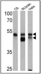
- Western blot analysis of pan Arrestin was performed by loading 25 µg of C6 (lane 1), rat brain (lane 2) and Hela (lane 3) cell lysates onto an SDS polyacrylamide gel. Proteins were transferred to a PVDF membrane and blocked at 4ºC overnight. The membrane was probed with a pan Arrestin polyclonal antibody (Product # PA1-730) at a dilution of 1:1000 overnight at 4°C, washed in TBST, and probed with an HRP-conjugated secondary antibody for 1 hr at room temperature in the dark. Chemiluminescent detection was performed using Pierce ECL Plus Western Blotting Substrate (Product # 32132). Results show a band at ~47-49 kDa.

Supportive validation
- Submitted by
- Invitrogen Antibodies (provider)
- Main image
- Experimental details
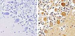
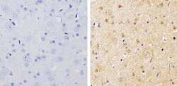
- Immunohistochemistry analysis of pan Arrestin showing staining in the cytoplasm and nucleus of paraffin-treated human cerebellum tissue (right) compared with a negative control in the absence of primary antibody (left). To expose target proteins, antigen retrieval was performed using 10mM sodium citrate (pH 6.0), microwaved for 8-15 min. Following antigen retrieval, tissues were blocked in 3% H2O2-methanol for 15 min at room temperature, washed with ddH2O and PBS, and then probed with a pan Arrestin polyclonal antibody (Product # PA1-730) diluted by 3% BSA-PBS at a dilution of 1:500 overnight at 4°C in a humidified chamber. Tissues were washed extensively in PBST and detection was performed using an HRP-conjugated secondary antibody followed by colorimetric detection using a DAB kit. Tissues were counterstained with hematoxylin and dehydrated with ethanol and xylene to prep for mounting.
- Submitted by
- Invitrogen Antibodies (provider)
- Main image
- Experimental details
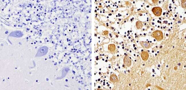
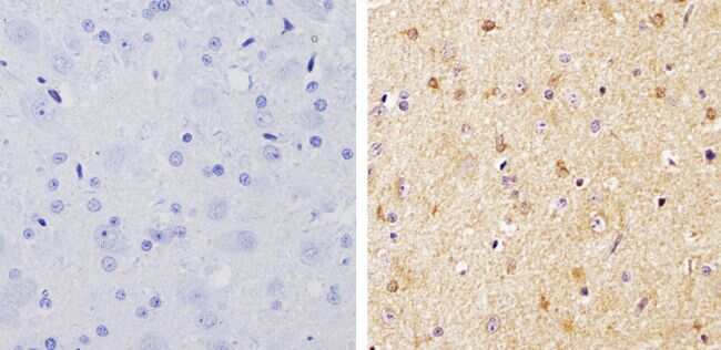
- Immunohistochemistry analysis of pan Arrestin showing staining in the cytoplasm and nucleus of paraffin-treated rat brain tissue (right) compared with a negative control in the absence of primary antibody (left). To expose target proteins, antigen retrieval was performed using 10mM sodium citrate (pH 6.0), microwaved for 8-15 min. Following antigen retrieval, tissues were blocked in 3% H2O2-methanol for 15 min at room temperature, washed with ddH2O and PBS, and then probed with a pan Arrestin polyclonal antibody (Product # PA1-730) diluted by 3% BSA-PBS at a dilution of 1:500 overnight at 4°C in a humidified chamber. Tissues were washed extensively in PBST and detection was performed using an HRP-conjugated secondary antibody followed by colorimetric detection using a DAB kit. Tissues were counterstained with hematoxylin and dehydrated with ethanol and xylene to prep for mounting.
- Submitted by
- Invitrogen Antibodies (provider)
- Main image
- Experimental details
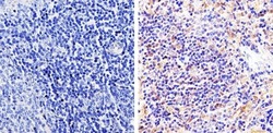
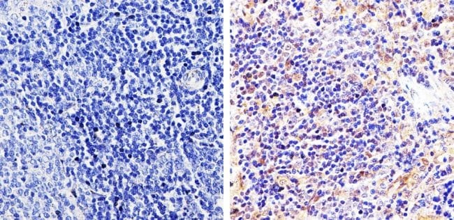
- Immunohistochemistry analysis of pan Arrestin showing staining in the cytoplasm and nucleus of paraffin-treated rat spleen tissue (right) compared with a negative control in the absence of primary antibody (left). To expose target proteins, antigen retrieval was performed using 10mM sodium citrate (pH 6.0), microwaved for 8-15 min. Following antigen retrieval, tissues were blocked in 3% H2O2-methanol for 15 min at room temperature, washed with ddH2O and PBS, and then probed with a pan Arrestin polyclonal antibody (Product # PA1-730) diluted by 3% BSA-PBS at a dilution of 1:500 overnight at 4°C in a humidified chamber. Tissues were washed extensively in PBST and detection was performed using an HRP-conjugated secondary antibody followed by colorimetric detection using a DAB kit. Tissues were counterstained with hematoxylin and dehydrated with ethanol and xylene to prep for mounting.
Supportive validation
- Submitted by
- Invitrogen Antibodies (provider)
- Main image
- Experimental details
- NULL