Explore
 Validate
 Learn
Learn Western blot
 Immunohistochemistry
Antibody data
- Antibody Data
- Antigen structure
- References [9]
- Comments [0]
- Validations
- Western blot [1]
- ELISA [1]
- Immunocytochemistry [1]
- Immunoprecipitation [1]
Submit
Validation data
Reference
Comment
Report error
- Product number
- H00060485-M02 - Provider product page

- Provider
- Novus Biologicals
- Proper citation
- Novus Cat#H00060485-M02, RRID:AB_547968
- Product name
- Mouse Monoclonal SAV1 Antibody
- Antibody type
- Monoclonal
- Description
- IgG purified. SAV1 - salvador homolog 1 (Drosophila)
- Reactivity
- Human, Mouse, Yeast
- Host
- Mouse
- Isotype
- IgG
- Vial size
- 0.1 mg
- Storage
- Aliquot and store at -20C or -80C. Avoid freeze-thaw cycles.
Submitted references FGF15 Activates Hippo Signaling to Suppress Bile Acid Metabolism and Liver Tumorigenesis.
Mammalian Hippo kinase pathway is downregulated by BCL-2 via protein degradation.
Kidney-specific knockout of Sav1 in the mouse promotes hyperproliferation of renal tubular epithelium through suppression of the Hippo pathway.
Hippo signaling mediates proliferation, invasiveness, and metastatic potential of clear cell renal cell carcinoma.
Spatial organization of Hippo signaling at the plasma membrane mediated by the tumor suppressor Merlin/NF2.
Mammalian ste20-like kinase and SAV1 promote 3T3-L1 adipocyte differentiation by activation of PPARγ.
Screening of binding proteins that interact with human Salvador 1 in a human fetal liver cDNA library by the yeast two-hybrid system.
LATS2 is a tumor suppressor gene of malignant mesothelioma.
Components of the Hippo pathway cooperate with Nek2 kinase to regulate centrosome disjunction.
Ji S, Liu Q, Zhang S, Chen Q, Wang C, Zhang W, Xiao C, Li Y, Nian C, Li J, Li J, Geng J, Hong L, Xie C, He Y, Chen X, Li X, Yin ZY, You H, Lin KH, Wu Q, Yu C, Johnson RL, Wang L, Chen L, Wang F, Zhou D
Developmental cell 2019 Feb 25;48(4):460-474.e9
Developmental cell 2019 Feb 25;48(4):460-474.e9
Mammalian Hippo kinase pathway is downregulated by BCL-2 via protein degradation.
Won GW, Park SH, Park J, Lee Y, Lee YH
Biochemical and biophysical research communications 2019 Apr 23;512(1):87-92
Biochemical and biophysical research communications 2019 Apr 23;512(1):87-92
Kidney-specific knockout of Sav1 in the mouse promotes hyperproliferation of renal tubular epithelium through suppression of the Hippo pathway.
Kai T, Tsukamoto Y, Hijiya N, Tokunaga A, Nakada C, Uchida T, Daa T, Iha H, Takahashi M, Nomura T, Sato F, Mimata H, Ikawa M, Seto M, Matsuura K, Moriyama M
The Journal of pathology 2016 May;239(1):97-108
The Journal of pathology 2016 May;239(1):97-108
Hippo signaling mediates proliferation, invasiveness, and metastatic potential of clear cell renal cell carcinoma.
Schütte U, Bisht S, Heukamp LC, Kebschull M, Florin A, Haarmann J, Hoffmann P, Bendas G, Buettner R, Brossart P, Feldmann G
Translational oncology 2014 Apr;7(2):309-21
Translational oncology 2014 Apr;7(2):309-21
Spatial organization of Hippo signaling at the plasma membrane mediated by the tumor suppressor Merlin/NF2.
Yin F, Yu J, Zheng Y, Chen Q, Zhang N, Pan D
Cell 2013 Sep 12;154(6):1342-55
Cell 2013 Sep 12;154(6):1342-55
Mammalian ste20-like kinase and SAV1 promote 3T3-L1 adipocyte differentiation by activation of PPARγ.
Park BH, Kim DS, Won GW, Jeon HJ, Oh BC, Lee Y, Kim EG, Lee YH
PloS one 2012;7(1):e30983
PloS one 2012;7(1):e30983
Screening of binding proteins that interact with human Salvador 1 in a human fetal liver cDNA library by the yeast two-hybrid system.
Li X, Luo X, Li Z, Wang G, Xiao H, Tao D, Gong J, Hu J
Molecular biology reports 2012 Aug;39(8):8225-30
Molecular biology reports 2012 Aug;39(8):8225-30
LATS2 is a tumor suppressor gene of malignant mesothelioma.
Murakami H, Mizuno T, Taniguchi T, Fujii M, Ishiguro F, Fukui T, Akatsuka S, Horio Y, Hida T, Kondo Y, Toyokuni S, Osada H, Sekido Y
Cancer research 2011 Feb 1;71(3):873-83
Cancer research 2011 Feb 1;71(3):873-83
Components of the Hippo pathway cooperate with Nek2 kinase to regulate centrosome disjunction.
Mardin BR, Lange C, Baxter JE, Hardy T, Scholz SR, Fry AM, Schiebel E
Nature cell biology 2010 Dec;12(12):1166-76
Nature cell biology 2010 Dec;12(12):1166-76
No comments: Submit comment
Supportive validation
- Submitted by
- Novus Biologicals (provider)
- Main image

- Experimental details
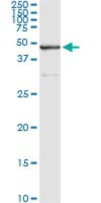
- Western Blot: SAV1 Antibody (3B2) [H00060485-M02] - SAV1 monoclonal antibody (M02), clone 3B2 Analysis of SAV1 expression in Hela S3 NE.

Supportive validation
- Submitted by
- Novus Biologicals (provider)
- Main image
- Experimental details
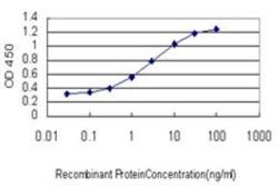
- ELISA: SAV1 Antibody (3B2) [H00060485-M02] - Detection limit for recombinant GST tagged SAV1 is approximately 0.1ng/ml as a capture antibody.

Supportive validation
- Submitted by
- Novus Biologicals (provider)
- Main image

- Experimental details
- Immunocytochemistry/Immunofluorescence: SAV1 Antibody (3B2) [H00060485-M02] - Analysis of monoclonal antibody to SAV1 on HeLa cell. Antibody concentration 60 ug/ml.

Supportive validation
- Submitted by
- Novus Biologicals (provider)
- Main image

- Experimental details
- Immunoprecipitation: SAV1 Antibody (3B2) [H00060485-M02] - Analysis of SAV1 transfected lysate using anti-SAV1 monoclonal antibody and Protein A Magnetic Bead, and immunoblotted with SAV1 MaxPab rabbit polyclonal antibody.