Explore
 Validate
 Learn
Learn Western blot
 Immunocytochemistry
Antibody data
- Antibody Data
- Antigen structure
- References [2]
- Comments [0]
- Validations
- Western blot [1]
- Immunocytochemistry [1]
- Immunohistochemistry [6]
Submit
Validation data
Reference
Comment
Report error
- Product number
- HPA011276 - Provider product page

- Provider
- Atlas Antibodies
- Proper citation
- Atlas Antibodies Cat#HPA011276, RRID:AB_1857503
- Product name
- Anti-SSR1
- Antibody type
- Polyclonal
- Reactivity
- Human, Mouse, Rat
- Host
- Rabbit
- Conjugate
- Unconjugated
- Antigen sequence
ESRKRKRPIQKVEMGTSSQNDVDMSWIPQETLNQI
NKASPRRLPRKRAQKRSVGSDE- Isotype
- IgG
- Vial size
- 100 µl
- Storage
- Store at +4°C for short term storage. Long time storage is recommended at -20°C.
Submitted references Architectural organization of the metabolic regulatory enzyme ghrelin O-acyltransferase.
Immunofluorescence and fluorescent-protein tagging show high correlation for protein localization in mammalian cells
Taylor MS, Ruch TR, Hsiao PY, Hwang Y, Zhang P, Dai L, Huang CRL, Berndsen CE, Kim MS, Pandey A, Wolberger C, Marmorstein R, Machamer C, Boeke JD, Cole PA
The Journal of biological chemistry 2013 Nov 8;288(45):32211-32228
The Journal of biological chemistry 2013 Nov 8;288(45):32211-32228
Immunofluorescence and fluorescent-protein tagging show high correlation for protein localization in mammalian cells
Stadler C, Rexhepaj E, Singan V, Murphy R, Pepperkok R, Uhlén M, Simpson J, Lundberg E
Nature Methods 2013 February;10(4):315-323
Nature Methods 2013 February;10(4):315-323
No comments: Submit comment
Enhanced validation
- Submitted by
- Atlas Antibodies (provider)
- Enhanced method
- Independent antibody validation
- Main image

- Experimental details
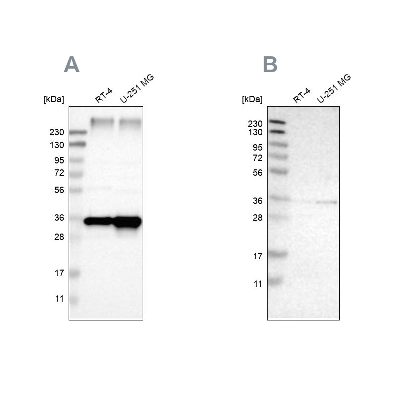
- Western blot analysis using Anti-SSR1 antibody HPA011276 (A) shows similar pattern to independent antibody HPA017062 (B).
Supportive validation
- Submitted by
- Atlas Antibodies (provider)
- Main image

- Experimental details
- Immunofluorescent staining of human cell line U-251 MG shows localization to endoplasmic reticulum.
- Sample type
- HUMAN

Enhanced validation
Supportive validation
- Submitted by
- Atlas Antibodies (provider)
- Enhanced method
- Independent antibody validation
- Main image

- Experimental details
- Immunohistochemical staining of human cerebral cortex, liver, lymph node and pancreas using Anti-SSR1 antibody HPA011276 (A) shows similar protein distribution across tissues to independent antibody HPA017062 (B).

Supportive validation
- Submitted by
- Atlas Antibodies (provider)
- Main image

- Experimental details
- Immunohistochemical staining of human gallbladder shows moderate cytoplasmic positivity in glandular cells.

- Submitted by
- Atlas Antibodies (provider)
- Main image

- Experimental details
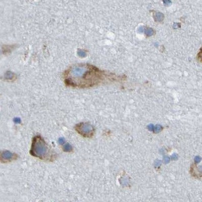
- Immunohistochemical staining of human cerebral cortex using Anti-SSR1 antibody HPA011276.
- Sample type
- HUMAN
- Submitted by
- Atlas Antibodies (provider)
- Main image
- Experimental details
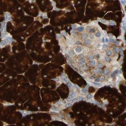
- Immunohistochemical staining of human pancreas using Anti-SSR1 antibody HPA011276.
- Sample type
- HUMAN

- Submitted by
- Atlas Antibodies (provider)
- Main image

- Experimental details
- Immunohistochemical staining of human liver using Anti-SSR1 antibody HPA011276.
- Sample type
- HUMAN

- Submitted by
- Atlas Antibodies (provider)
- Main image

- Experimental details
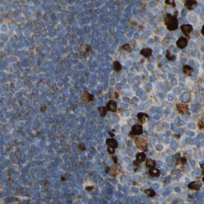
- Immunohistochemical staining of human lymph node using Anti-SSR1 antibody HPA011276.
- Sample type
- HUMAN