 Explore
 Validate
 Learn
Learn Western blot
 Immunocytochemistry
Antibody data
- Antibody Data
- Antigen structure
- References [1]
- Comments [0]
- Validations
- Immunocytochemistry [1]
- Immunohistochemistry [1]
Submit
Validation data
Reference
Comment
Report error
- Product number
- GTX22798 - Provider product page

- Provider
- GeneTex
- Proper citation
- GeneTex Cat#GTX22798, RRID:AB_368880
- Product name
- Calnexin antibody [AF18]
- Antibody type
- Monoclonal
- Reactivity
- Human, Mouse, Rat, Simian
- Host
- Mouse
Submitted references Comparison of five commercial extraction kits for subsequent membrane protein profiling.
Bünger S, Roblick UJ, Habermann JK
Cytotechnology 2009 Dec;61(3):153-9
Cytotechnology 2009 Dec;61(3):153-9
No comments: Submit comment
Supportive validation
- Submitted by
- GeneTex (provider)
- Main image
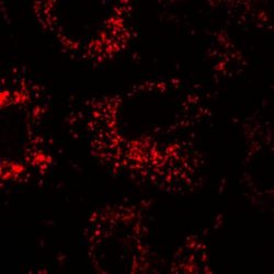
- Experimental details
- Immunocytochemical analysis of Calnexin in COS7 cells. Transfected COS7 cells were blocked in 1% BSA, 5% normal goat serum, and 0.1% Triton X-100 in 1X PBS, and then stained (1:500), followed by a fluorophore-conjugated secondary antibody (red). Magnification = 100X.

Supportive validation
- Submitted by
- GeneTex (provider)
- Main image

- Experimental details
- Immunohistochemistry was performed on cancer biopsies of deparaffinized human hepatocarcinoma tissues. To expose target proteins, heat induced antigen retrieval was performed using 10mM sodium citrate (pH6.0) buffer, microwaved for 8-15 minutes. Following antigen retrieval tissues were blocked in 3% BSA-PBS for 30 minutes at room temperature. Tissues were then probed at a dilution of 1:20 with or without Calnexin antibody [AF18] overnight at 4¢XC in a humidified chamber. Tissues were washed extensively with PBST and endogenous peroxidase activity was quenched with a peroxidase suppressor. Detection was performed using a biotin-conjugated secondary antibody and SA-HRP, followed by colorimetric detection using DAB. Tissues were counterstained with hematoxylin and prepped for mounting.
