Explore
 Validate
 Learn
Learn Western blot
 Immunocytochemistry
Antibody data
- Antibody Data
- Antigen structure
- References [1]
- Comments [0]
- Validations
- Western blot [1]
- Immunohistochemistry [5]
- Flow cytometry [2]
Submit
Validation data
Reference
Comment
Report error
- Product number
- NBP2-36570 - Provider product page

- Provider
- Novus Biologicals
- Product name
- Mouse Monoclonal Calnexin Antibody
- Antibody type
- Monoclonal
- Description
- Protein G purified.
- Reactivity
- Human
- Host
- Mouse
- Isotype
- IgG
- Vial size
- 0.1 mg
- Concentration
- 1.0 mg/ml
- Storage
- Store at 4C short term. Aliquot and store at -20C long term. Avoid freeze-thaw cycles.
Submitted references A dendritic cell receptor-targeted chimeric immunotherapeutic protein (C-HBV) for the treatment of chronic hepatitis B.
Ma A, Motyka B, Gutfreund K, Shi YE, George R
Human vaccines & immunotherapeutics 2020 Apr 2;16(4):756-778
Human vaccines & immunotherapeutics 2020 Apr 2;16(4):756-778
No comments: Submit comment
Supportive validation
- Submitted by
- Novus Biologicals (provider)
- Main image

- Experimental details
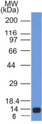
- Western Blot: Calnexin Antibody (1C2.2D11) [NBP2-36570] - Analysis of 11 kDa Partial Recombinant Human Calnexin protein with Calnexin antibody (clone 1C2.2D11) at 0.5 ug/mL.
Supportive validation
- Submitted by
- Novus Biologicals (provider)
- Main image
- Experimental details
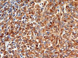
- Immunohistochemistry-Paraffin: Calnexin Antibody (1C2.2D11) [NBP2-36570] - Analysis of FFPE tissue section of malignant stromal tumor of the human small bowel using mouse monoclonal Calnexin antibody (clone 1C2.2D11) at 7 ug/mL concentration. The cancer cells showed a very strong cytoplasmic reactivity for Calnexin.
- Submitted by
- Novus Biologicals (provider)
- Main image

- Experimental details
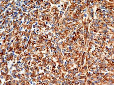
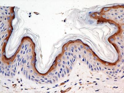
- Immunohistochemistry-Paraffin: Calnexin Antibody (1C2.2D11) [NBP2-36570] - Analysis of FFPE tissue section of human skin using mouse monoclonal Calnexin antibody (clone 1C2.2D11) at 7 ug/mL concentration. The outermost keratinocytes layer of the epidermis showed cytoplasmic positivity for Calnexin protein.
- Submitted by
- Novus Biologicals (provider)
- Main image

- Experimental details
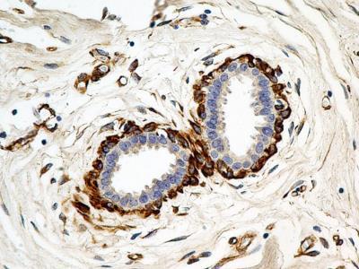
- Immunohistochemistry-Paraffin: Calnexin Antibody (1C2.2D11) [NBP2-36570] - Analysis of FFPE tissue section of normal human breast using mouse monoclonal Calnexin antibody (clone 1C2.2D11) at 7 ug/mL concentration. The myoepithelial cells around the lobules depicted a very strong cytoplasmic staining.
- Submitted by
- Novus Biologicals (provider)
- Main image

- Experimental details
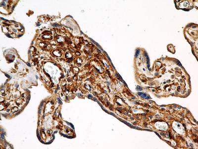
- Immunohistochemistry-Paraffin: Calnexin Antibody (1C2.2D11) [NBP2-36570] - Analysis of FFPE tissue section of human placenta using mouse monoclonal Calnexin antibody (clone 1C2.2D11) at 7 ug/mL concentration.
- Submitted by
- Novus Biologicals (provider)
- Main image

- Experimental details
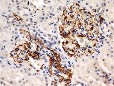
- Immunohistochemistry-Paraffin: Calnexin Antibody (1C2.2D11) [NBP2-36570] - Analysis of FFPE tissue section of normal human kidney using mouse monoclonal Calnexin antibody (clone 1C2.2D11) at 7 ug/mL concentration. The cells of Glomeruli developed strong cytoplasmic staining.
Supportive validation
- Submitted by
- Novus Biologicals (provider)
- Main image

- Experimental details
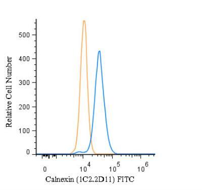
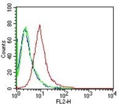
- Flow (Intracellular): Calnexin Antibody (1C2.2D11) [NBP2-36570] - Intracellular staining of human Calnexin in Flow cytometry using 5.0 ug of antibody per 1 million cells. Isotype control was mouse IgG2b kappa.
- Submitted by
- Novus Biologicals (provider)
- Main image

- Experimental details
- Flow (Intracellular): Calnexin Antibody (1C2.2D11) [NBP2-36570] - An intracellular stain was performed on HeLa cells with Calnexin Antibody (1C2.2D11) NBP2-36570F (blue) and a matched isotype control (orange). Cells were fixed with 4% PFA and then permeabilized with 0.1% saponin. Cells were incubated in an antibody dilution of 10 ug/mL for 30 minutes at room temperature. Both antibodies were conjugated to FITC.