Explore
 Validate
 Learn
Learn Western blot
 Immunocytochemistry
Antibody data
- Antibody Data
- Antigen structure
- References [6]
- Comments [0]
- Validations
- Western blot [3]
- Immunohistochemistry [1]
- Flow cytometry [2]
Submit
Validation data
Reference
Comment
Report error
- Product number
- NB100-1974 - Provider product page

- Provider
- Novus Biologicals
- Proper citation
- Novus Cat#NB100-1974, RRID:AB_10001873
- Product name
- Rabbit Polyclonal Calnexin Antibody
- Antibody type
- Polyclonal
- Description
- Immunogen affinity purified.
- Reactivity
- Human, Mouse, Rat, Hamster
- Host
- Rabbit
- Isotype
- IgG
- Vial size
- 0.1 ml
- Concentration
- 1 mg/ml
- Storage
- Store at 4C short term. Aliquot and store at -20C long term. Avoid freeze-thaw cycles.
Submitted references Exosomal hsa-miR199a-3p Promotes Proliferation and Migration in Neuroblastoma.
Interplay between ChREBP and SREBP-1c coordinates postprandial glycolysis and lipogenesis in livers of mice.
Glutathione S-Transferase P-Mediated Protein S-Glutathionylation of Resident Endoplasmic Reticulum Proteins Influences Sensitivity to Drug-Induced Unfolded Protein Response.
The HMGB1/RAGE inflammatory pathway promotes pancreatic tumor growth by regulating mitochondrial bioenergetics.
Stabilization of the μ-opioid receptor by truncated single transmembrane splice variants through a chaperone-like action.
Characterization of zebrafish von Willebrand factor reveals conservation of domain structure, multimerization, and intracellular storage.
Ma J, Xu M, Yin M, Hong J, Chen H, Gao Y, Xie C, Shen N, Gu S, Mo X
Frontiers in oncology 2019;9:459
Frontiers in oncology 2019;9:459
Interplay between ChREBP and SREBP-1c coordinates postprandial glycolysis and lipogenesis in livers of mice.
Linden AG, Li S, Choi HY, Fang F, Fukasawa M, Uyeda K, Hammer RE, Horton JD, Engelking LJ, Liang G
Journal of lipid research 2018 Mar;59(3):475-487
Journal of lipid research 2018 Mar;59(3):475-487
Glutathione S-Transferase P-Mediated Protein S-Glutathionylation of Resident Endoplasmic Reticulum Proteins Influences Sensitivity to Drug-Induced Unfolded Protein Response.
Ye ZW, Zhang J, Ancrum T, Manevich Y, Townsend DM, Tew KD
Antioxidants & redox signaling 2017 Feb 20;26(6):247-261
Antioxidants & redox signaling 2017 Feb 20;26(6):247-261
The HMGB1/RAGE inflammatory pathway promotes pancreatic tumor growth by regulating mitochondrial bioenergetics.
Kang R, Tang D, Schapiro NE, Loux T, Livesey KM, Billiar TR, Wang H, Van Houten B, Lotze MT, Zeh HJ
Oncogene 2014 Jan 30;33(5):567-77
Oncogene 2014 Jan 30;33(5):567-77
Stabilization of the μ-opioid receptor by truncated single transmembrane splice variants through a chaperone-like action.
Xu J, Xu M, Brown T, Rossi GC, Hurd YL, Inturrisi CE, Pasternak GW, Pan YX
The Journal of biological chemistry 2013 Jul 19;288(29):21211-27
The Journal of biological chemistry 2013 Jul 19;288(29):21211-27
Characterization of zebrafish von Willebrand factor reveals conservation of domain structure, multimerization, and intracellular storage.
Ghosh A, Vo A, Twiss BK, Kretz CA, Jozwiak MA, Montgomery RR, Shavit JA
Advances in hematology 2012;2012:214209
Advances in hematology 2012;2012:214209
No comments: Submit comment
Supportive validation
- Submitted by
- Novus Biologicals (provider)
- Main image
- Experimental details
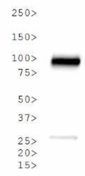
- Western Blot: Calnexin Antibody [NB100-1974] - Analysis of Calnexin in HeLa whole cell lysate.
- Submitted by
- Novus Biologicals (provider)
- Main image

- Experimental details
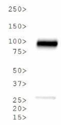
- Simple Western: Calnexin Antibody [NB100-1974] - Image shows a specific band for Calnexin in 0.1 mg/mL of HeLa lysate. This experiment was performed under reducing conditions using the 12-230 kDa separation system.

- Submitted by
- Novus Biologicals (provider)
- Main image
- Experimental details
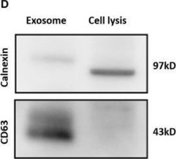
- Western Blot: Calnexin Antibody [NB100-1974] - Identification of plasma exosomes and differentially expressed exosomal miRNAs. Confirmation of the exosomes markers with Western blotting indicated the presence of CD63 but the absence of calnexin in exosomes. Image collected and cropped by CiteAb from the following publication (https://www.frontiersin.org/article/10.3389/fonc.2019.00459/full), licensed under a CC-BY licence.

Supportive validation
- Submitted by
- Novus Biologicals (provider)
- Main image

- Experimental details
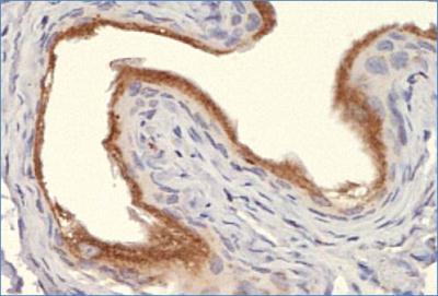
- Immunohistochemistry: Calnexin Antibody [NB100-1974] - Analysis of Calnexin in mouse bladder using DAB with hematoxylin counterstain.
Supportive validation
- Submitted by
- Novus Biologicals (provider)
- Main image

- Experimental details
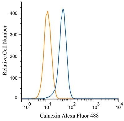
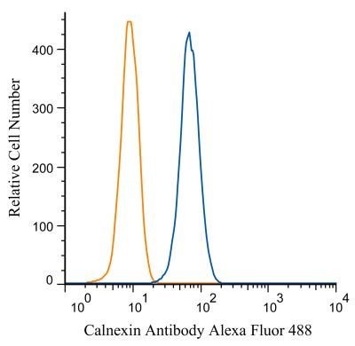
- Flow Cytometry: Calnexin Antibody [NB100-1974] - An intracellular stain was performed on HeLa cells with Calnexin antibody NB100-1974AF488 (blue) and a matched isotype control NBP2-24893AF488 (orange). Cells were fixed with 4% PFA and then permeablized with 0.1% saponin. Cells were incubated in an antibody dilution of 5 ug/mL for 30 minutes at room temperature. Both antibodies were conjugated to Alexa Fluor 488. Image using the Alexa Fluor 488 form of this antibody.
- Submitted by
- Novus Biologicals (provider)
- Main image

- Experimental details
- Flow Cytometry: Calnexin Antibody [NB100-1974] - Analysis of Alexa Fluor (R) 488 conjugate of NB100-1974. An intracellular stain was performed on Jurkat cells with Calnexin antibody NB100-1974AF488 (blue) and a matched isotype control NBP2-24893AF488 (orange).