Explore
 Validate
 Learn
Learn Western blot
Antibody data
- Antibody Data
- Antigen structure
- References [2]
- Comments [0]
- Validations
- Western blot [3]
- Immunocytochemistry [2]
- Immunohistochemistry [1]
- Other assay [2]
Submit
Validation data
Reference
Comment
Report error
- Product number
- PA5-19169 - Provider product page

- Provider
- Invitrogen Antibodies
- Product name
- Calnexin Polyclonal Antibody
- Antibody type
- Polyclonal
- Antigen
- Synthetic peptide
- Description
- This antibody is predicted to react with bovine, canine, porcine and rat based on sequence homology. This antibody is tested in Peptide ELISA: antibody detection limit dilution 128,000.
- Reactivity
- Human
- Host
- Goat
- Isotype
- IgG
- Vial size
- 100 µg
- Concentration
- 0.5 mg/mL
- Storage
- -20° C, Avoid Freeze/Thaw Cycles
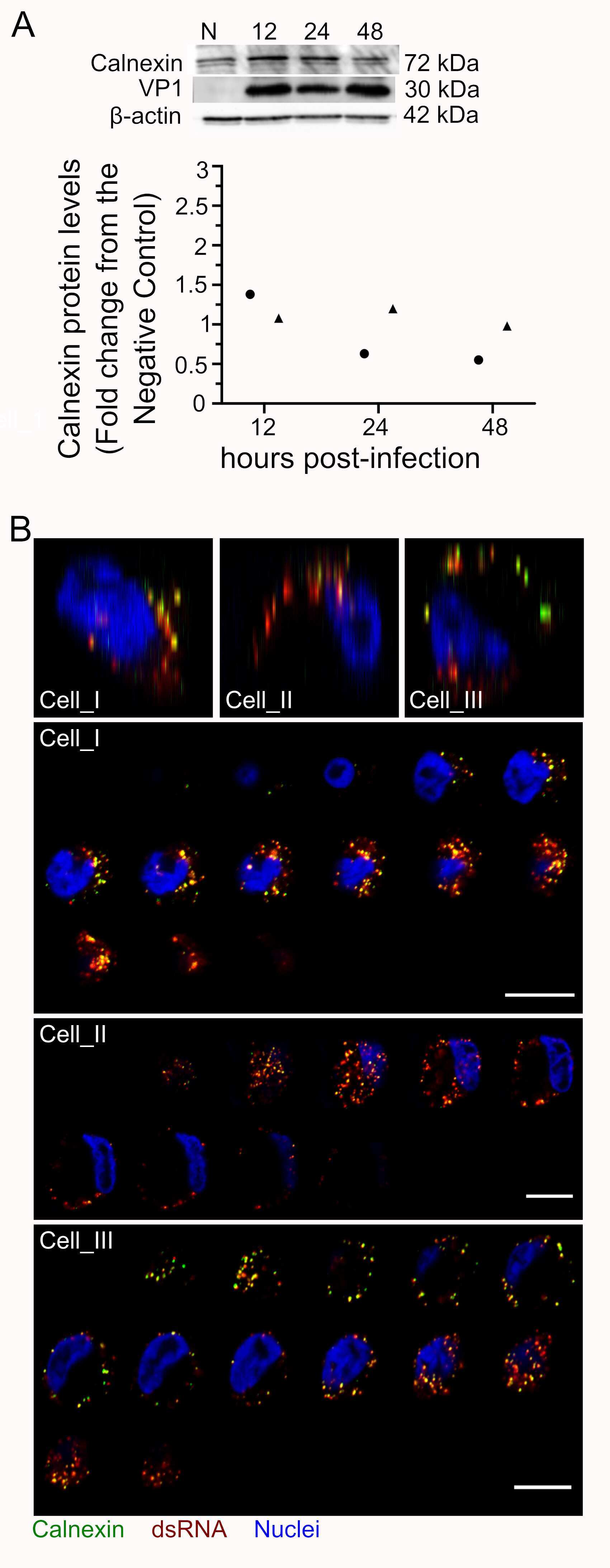
Submitted references Rhinovirus C replication is associated with the endoplasmic reticulum and triggers cytopathic effects in an in vitro model of human airway epithelium.
Ubiquitination of G3BP1 mediates stress granule disassembly in a context-specific manner.
Gagliardi TB, Goldstein ME, Song D, Gray KM, Jung JW, Ignacio MA, Stroka KM, Duncan GA, Scull MA
PLoS pathogens 2022 Jan;18(1):e1010159
PLoS pathogens 2022 Jan;18(1):e1010159
Ubiquitination of G3BP1 mediates stress granule disassembly in a context-specific manner.
Gwon Y, Maxwell BA, Kolaitis RM, Zhang P, Kim HJ, Taylor JP
Science (New York, N.Y.) 2021 Jun 25;372(6549):eabf6548
Science (New York, N.Y.) 2021 Jun 25;372(6549):eabf6548
No comments: Submit comment
Supportive validation
- Submitted by
- Invitrogen Antibodies (provider)
- Main image

- Experimental details
- Western Blot staining of Mouse Heart lysate using Product # PA5-19169 at a concentration of 0.3 µg/mL, the primary antibody incubation was 1 hour and the detection method was chemiluminescence.

- Submitted by
- Invitrogen Antibodies (provider)
- Main image

- Experimental details
- Western blot analysis was performed on whole cell extracts (30 µg lysate) of HeLa (Lane 1), HCT116 (Lane 2), A431 (Lane 3), HepG2 (Lane 4), Raji (Lane 5), MOLT-4 (Lane 6), Jurkat (Lane 7) and RAW 264.7 (Lane 8). The blot was probed with anti-Calnexin Polyclonal Antibody (Product # PA5-19169, 1:3000 dilution) and detected by chemiluminescence using Rabbit anti-Goat IgG (H+L) Superclonal™ Secondary Antibody, HRP conjugate (Product # A27014, 0.25 µg/mL, 1:4000 dilution). A 72 kDa band corresponding to Calnexin was observed across all the cell lines tested.

- Submitted by
- Invitrogen Antibodies (provider)
- Main image

- Experimental details
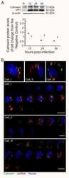
- Knockdown of Calnexin was achieved by transfecting HeLa cells with Calnexin specific siRNAs (Silencer® select Product # s2376). Western blot analysis (Fig. a) was performed using whole cell extracts from the Calnexin knockdown cells (lane 3), non-specific scrambled siRNA transfected cells (lane 2) and untransfected cells (lane 1). The blot was probed with Calnexin Polyclonal Antibody (Product # PA5-19169, 1µg/ml) and Goat anti-Rabbit IgG (H+L) Superclonal™ Secondary Antibody, HRP conjugate (Product # A27036, 0.25µg/ml, 1:4000 dilution). Densitometric analysis of this western blot is shown in histogram (Fig. b). Decrease in signal upon siRNA mediated knock down confirms that antibody is specific to Calnexin.

Supportive validation
- Submitted by
- Invitrogen Antibodies (provider)
- Main image
- Experimental details
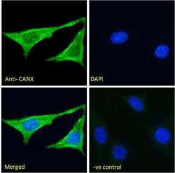
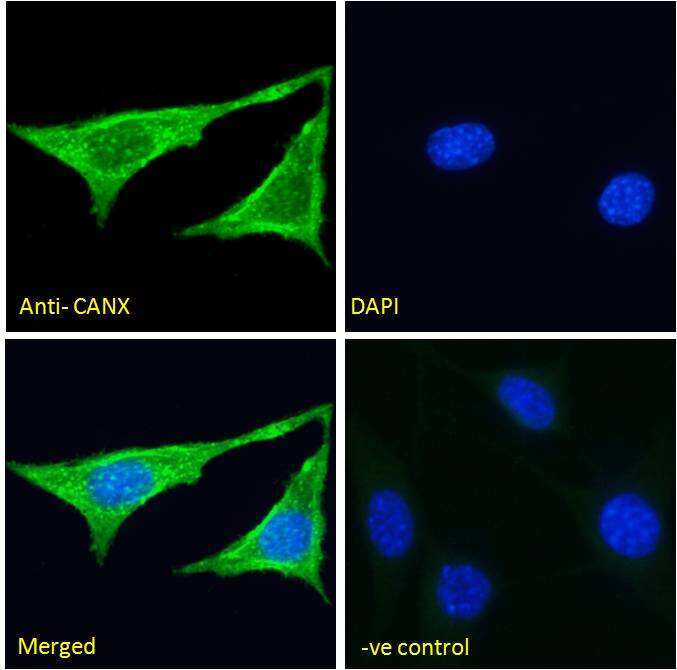
- Immunofluorescence analysis of Calnexin in NIH3T3 cells using a Calnexin monoclonal antibody (Product # PA5-19169) at 5 µg/mL for1hr. The cells were paraformaldehyde fixed and permeabilized with 0.15% Triton. Primary incubation was followed by Alexa Fluor 488 secondary antibody (2 µg/mL) showing cytoplasmic staining. The nuclear stain is DAPI (blue). Negative control: Unimmunized goat IgG (5 µg/mL)followed by Alexa Fluor 488 secondary antibody (2 µg/mL).
- Submitted by
- Invitrogen Antibodies (provider)
- Main image
- Experimental details
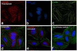
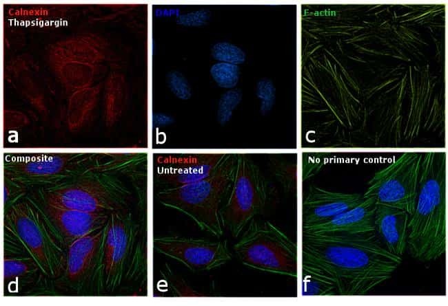
- Immunofluorescence analysis of Calnexin was performed using 70% confluent log phase HeLa cells treated with Thapsigargin (1uM for 24hrs). The cells were fixed with 4% paraformaldehyde for 10 minutes, permeabilized with 0.1% Triton™ X-100 for 15 minutes, and blocked with 1% BSA for 1 hour at room temperature. The cells were labeled with Calnexin Polyclonal Antibody (Product # PA5-19169) at 1:200 dilution in 0.1% BSA, incubated at 4 degree celsius overnight and then labeled with Rabbit anti-Goat IgG (H+L) Superclonal™ Secondary Antibody, Alexa Fluor 594 conjugate (Product # A27016) at a dilution of 1:2000 for 45 minutes at room temperature (Panel a: red). Nuclei (Panel b: blue) were stained with ProLong™ Diamond Antifade Mountant with DAPI (Product # P36962). F-actin (Panel c: green) was stained with Alexa Fluor™ 488 Phalloidin (Product # A12379, 1:300). Panel d represents the merged image showing Calnexin in the ER and cytoplasm. Panel e represents the untreated cells showing lower expression levels. Panel f represents control cells with no primary antibody to assess background. The images were captured at 60X magnification.
Supportive validation
- Submitted by
- Invitrogen Antibodies (provider)
- Main image

- Experimental details
- Immunohistochemical analysis of Calnexin in paraffin embedded human kidney using a Calnexin polyclonal antibody (Product #PA5-19169) at a concentration of 3.8 µg/mL. Steamed antigen retrieval was performed with pH 6 buffered citrate. Samples were then stained with alkaline phosphatase.

Supportive validation
- Submitted by
- Invitrogen Antibodies (provider)
- Main image
- Experimental details
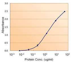
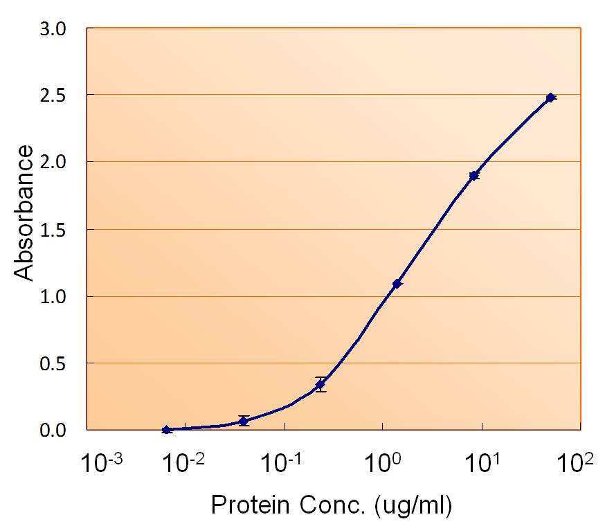
- ELISA of Calnexin using two Calnexin antibodies. The reporter antibody (Product # PA5-19169) was used at a concentration of 1.5 µg/ml and the reporter antibody was used at a concentration of 2.5 µg/ml.
- Submitted by
- Invitrogen Antibodies (provider)
- Main image
- Experimental details
- NULL