 Explore
 Validate
 Learn
Learn Western blot
Antibody data
- Antibody Data
- Antigen structure
- References [3]
- Comments [0]
- Validations
- Western blot [6]
- Immunocytochemistry [2]
Submit
Validation data
Reference
Comment
Report error
- Product number
- GTX122674 - Provider product page

- Provider
- GeneTex
- Proper citation
- GeneTex Cat#GTX122674, RRID:AB_11177321
- Product name
- ASGPR1 antibody [N1C3]
- Antibody type
- Polyclonal
- Reactivity
- Human, Mouse
- Host
- Rabbit
Submitted references Circulating extracellular vesicles with specific proteome and liver microRNAs are potential biomarkers for liver injury in experimental fatty liver disease.
Intracellular delivery of cytochrome c by galactosylated albumin to hepatocarcinoma cells.
Effect of high glucose on secreted proteome in cultured retinal pigmented epithelium cells: its possible relevance to clinical diabetic retinopathy.
Povero D, Eguchi A, Li H, Johnson CD, Papouchado BG, Wree A, Messer K, Feldstein AE
PloS one 2014;9(12):e113651
PloS one 2014;9(12):e113651
Intracellular delivery of cytochrome c by galactosylated albumin to hepatocarcinoma cells.
Yeh TH, Wu FL, Shen LJ
Journal of drug targeting 2014 Jul;22(6):528-35
Journal of drug targeting 2014 Jul;22(6):528-35
Effect of high glucose on secreted proteome in cultured retinal pigmented epithelium cells: its possible relevance to clinical diabetic retinopathy.
Chen YH, Chou HC, Lin ST, Chen YW, Lo YW, Chan HL
Journal of proteomics 2012 Dec 21;77:111-28
Journal of proteomics 2012 Dec 21;77:111-28
No comments: Submit comment
Supportive validation
- Submitted by
- GeneTex (provider)
- Main image

- Experimental details
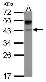
- Sample (30 ?g of whole cell lysate) A: HepG2 12% SDS PAGE GTX122674 diluted at 1:3000 The HRP-conjugated anti-rabbit IgG antibody (GTX213110-01) was used to detect the primary antibody.
- Submitted by
- GeneTex (provider)
- Main image

- Experimental details
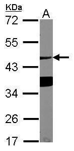
- Sample (30 ?g of whole cell lysate) A: NIH-3T3 12% SDS PAGE GTX122674 diluted at 1:1000 The HRP-conjugated anti-rabbit IgG antibody (GTX213110-01) was used to detect the primary antibody.
- Submitted by
- GeneTex (provider)
- Main image
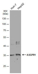
- Experimental details
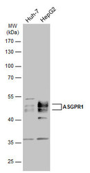
- Various whole cell extracts (30 ?g) were separated by 10% SDS-PAGE, and the membrane was blotted with ASGPR1 antibody [N1C3] (GTX122674) diluted at 1:3000. The HRP-conjugated anti-rabbit IgG antibody (GTX213110-01) was used to detect the primary antibody.
- Submitted by
- GeneTex (provider)
- Main image
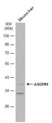
- Experimental details
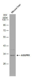
- Mouse tissue extract (50 ?g) was separated by 10% SDS-PAGE, and the membrane was blotted with ASGPR1 antibody [N1C3] (GTX122674) diluted at 1:3000. The HRP-conjugated anti-rabbit IgG antibody (GTX213110-01) was used to detect the primary antibody.
- Submitted by
- GeneTex (provider)
- Main image
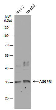
- Experimental details
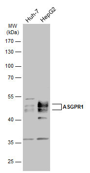
- Various whole cell extracts (30 ?g) were separated by 10% SDS-PAGE, and the membrane was blotted with ASGPR1 antibody [N1C3] (GTX122674) diluted at 1:3000. The HRP-conjugated anti-rabbit IgG antibody (GTX213110-01) was used to detect the primary antibody.
- Submitted by
- GeneTex (provider)
- Main image
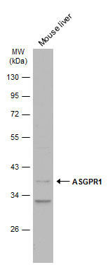
- Experimental details
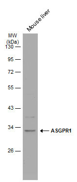
- Mouse tissue extract (50 ?g) was separated by 10% SDS-PAGE, and the membrane was blotted with ASGPR1 antibody [N1C3] (GTX122674) diluted at 1:3000. The HRP-conjugated anti-rabbit IgG antibody (GTX213110-01) was used to detect the primary antibody.
Supportive validation
- Submitted by
- GeneTex (provider)
- Main image
- Experimental details
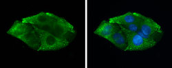
- ASGPR1 antibody [N1C3] detects secreted ASGPR1 protein by immunofluorescent analysis.Sample: HepG2 cells were fixed in 4% paraformaldehyde at RT for 15 min.Green: ASGPR1 protein stained by ASGPR1 antibody [N1C3] (GTX122674) diluted at 1:500.Blue: Hoechst 33342 staining.

- Submitted by
- GeneTex (provider)
- Main image
- Experimental details
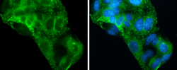
- ASGPR1 antibody [N1C3] detects secreted ASGPR1 protein by immunofluorescent analysis.Sample: HepG2 cells were fixed in 4% paraformaldehyde at RT for 15 min.Green: ASGPR1 stained by ASGPR1 antibody [N1C3] (GTX122674) diluted at 1:500.Blue: Hoechst 33342 staining.
