 Explore
 Validate
 Learn
Learn Western blot
 Immunoprecipitation
Antibody data
- Antibody Data
- Antigen structure
- References [0]
- Comments [0]
- Validations
- Western blot [2]
- Immunohistochemistry [2]
Submit
Validation data
Reference
Comment
Report error
- Product number
- PA5-72899 - Provider product page

- Provider
- Invitrogen Antibodies
- Product name
- BFAR Polyclonal Antibody
- Antibody type
- Polyclonal
- Antigen
- Synthetic peptide
- Reactivity
- Human
- Host
- Rabbit
- Isotype
- IgG
- Vial size
- 50 µL
- Concentration
- Conc. Not Determined
- Storage
- Store at 4°C short term. For long term storage, store at -20°C, avoiding freeze/thaw cycles.
No comments: Submit comment
Supportive validation
- Submitted by
- Invitrogen Antibodies (provider)
- Main image

- Experimental details
- Western blot analysis of BFAR in human brain, heart and mouse liver tissue lysate using 25 µg of lysate. Sample was incubated with BFAR polyclonal antibody (Product # PA5-72899).

- Submitted by
- Invitrogen Antibodies (provider)
- Main image
- Experimental details
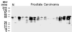
- Western blot analysis of BFAR in 25 µg/lane normal prostate and prostate carcinoma tissue lysates. Samples were incubated in BFAR polyclonal antibody (Product # PA5-72899). Tissue lysates from 15 different prostate carcinoma patients show variable expression of BAR with respect to banding patterns and amount of BAR expression. Major BAR bands typically migrate as a single band or as a doublet. These bands are typically observed at ~50-54 kDa. This is higher than the predicted molecular weight from the 450 amino acid BAR sequence, and may represent phosphorylation or other post-translational modifications. N = tissue lysate from normal prostate.

Supportive validation
- Submitted by
- Invitrogen Antibodies (provider)
- Main image

- Experimental details
- Immunohistochemical analysis of BFAR in formalin-fixed, paraffin-embedded tissue section. Samples were incubated in BFAR polyclonal antibody (Product # PA5-72899) using a dilution of 1:2000. A. Two cores from a human gliobastoma tissue microarray: 1 = fibrillary astrocytoma (grade I), and 2 = anaplastic glioma (grade III). B. Higher magnification from the fibrillary astrocytoma (shown in A). C. Higher magnification from the anaplastic glioma (shown in A). D. Normal human brain striatum with positive medium spiny neurons, the major neuronal cell type of the striatum. Hematoxylin-eosin counterstain.

- Submitted by
- Invitrogen Antibodies (provider)
- Main image
- Experimental details
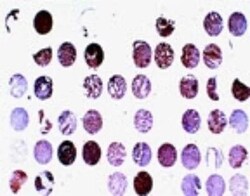
- Immunohistochemical analysis of BFAR in formalin-fixed, paraffin-embedded human prostate carcinoma tissue array. Samples were incubated in BFAR polyclonal antibody (Product # PA5-72899) using a dilution of 1:2000. Hematoxylin-eosin counterstain. Variable BAR expression is seen between patient samples.
