 Explore
 Validate
 Learn
Learn Western blot
 Immunocytochemistry
Antibody data
- Antibody Data
- Antigen structure
- References [4]
- Comments [0]
- Validations
- Western blot [4]
- Immunohistochemistry [1]
Submit
Validation data
Reference
Comment
Report error
- Product number
- NBP1-86589 - Provider product page

- Provider
- Novus Biologicals
- Proper citation
- Novus Cat#NBP1-86589, RRID:AB_11016077
- Product name
- Rabbit Polyclonal TRNT1 Antibody
- Antibody type
- Polyclonal
- Description
- Immunogen affinity purified. Specificity of human TRNT1 antibody verified on a Protein Array containing target protein plus 383 other non-specific proteins.
- Reactivity
- Human, Rat
- Host
- Rabbit
- Isotype
- IgG
- Vial size
- 0.1 ml
- Storage
- Store at 4C short term. Aliquot and store at -20C long term. Avoid freeze-thaw cycles.
Submitted references ELAC1 Repairs tRNAs Cleaved during Ribosome-Associated Quality Control.
Mechanism for recycling tRNAs on stalled ribosomes.
Hypomorphic mutations in TRNT1 cause retinitis pigmentosa with erythrocytic microcytosis.
Immunofluorescence and fluorescent-protein tagging show high correlation for protein localization in mammalian cells.
Yip MCJ, Savickas S, Gygi SP, Shao S
Cell reports 2020 Feb 18;30(7):2106-2114.e5
Cell reports 2020 Feb 18;30(7):2106-2114.e5
Mechanism for recycling tRNAs on stalled ribosomes.
Yip MCJ, Keszei AFA, Feng Q, Chu V, McKenna MJ, Shao S
Nature structural & molecular biology 2019 May;26(5):343-349
Nature structural & molecular biology 2019 May;26(5):343-349
Hypomorphic mutations in TRNT1 cause retinitis pigmentosa with erythrocytic microcytosis.
DeLuca AP, Whitmore SS, Barnes J, Sharma TP, Westfall TA, Scott CA, Weed MC, Wiley JS, Wiley LA, Johnston RM, Schnieders MJ, Lentz SR, Tucker BA, Mullins RF, Scheetz TE, Stone EM, Slusarski DC
Human molecular genetics 2016 Jan 1;25(1):44-56
Human molecular genetics 2016 Jan 1;25(1):44-56
Immunofluorescence and fluorescent-protein tagging show high correlation for protein localization in mammalian cells.
Stadler C, Rexhepaj E, Singan VR, Murphy RF, Pepperkok R, Uhlén M, Simpson JC, Lundberg E
Nature methods 2013 Apr;10(4):315-23
Nature methods 2013 Apr;10(4):315-23
No comments: Submit comment
Supportive validation
- Submitted by
- Novus Biologicals (provider)
- Main image

- Experimental details
- Simple Western: TRNT1 Antibody [NBP1-86589] - Simple Western lane view shows a specific band for TRNT1 in 0.2 mg/ml of RT-4 (Left) and U-251MG (Right) lysate. This experiment was performed under reducing conditions using the 12-230 kDa separation system.

- Submitted by
- Novus Biologicals (provider)
- Main image

- Experimental details
- Western Blot: TRNT1 Antibody [NBP1-86589] - Analysis in mouse cell line NIH-3T3 and rat cell line NBT-II.

- Submitted by
- Novus Biologicals (provider)
- Main image

- Experimental details
- Western Blot: TRNT1 Antibody [NBP1-86589] - Analysis in human cell line A-549.

- Submitted by
- Novus Biologicals (provider)
- Main image
- Experimental details
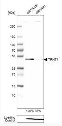
- Western Blot: TRNT1 Antibody [NBP1-86589] - Analysis in A-549 cells transfected with control siRNA, target specific siRNA probe #1, using Anti-TRNT1 antibody. Remaining relative intensity is presented. Loading control: Anti-PPIB.

Supportive validation
- Submitted by
- Novus Biologicals (provider)
- Main image

- Experimental details
- Immunohistochemistry-Paraffin: TRNT1 Antibody [NBP1-86589] - Staining of human stomach shows moderate cytoplasmic and nuclear positivity in glandular cells.
