 Explore
 Validate
 Learn
Learn Western blot
 ELISA
ELISAAntibody data
- Antibody Data
- Antigen structure
- References [0]
- Comments [0]
- Validations
- Western blot [2]
Submit
Validation data
Reference
Comment
Report error
- Product number
- NB600-470 - Provider product page

- Provider
- Novus Biologicals
- Proper citation
- Novus Cat#NB600-470, RRID:AB_10001258
- Product name
- Rabbit Polyclonal UBL5 Antibody
- Antibody type
- Polyclonal
- Description
- Ion exchange chromatography. Hub 1
- Reactivity
- Yeast
- Host
- Rabbit
- Isotype
- IgG
- Vial size
- 0.5 mg
- Concentration
- LYOPH
- Storage
- Store lyophilized antibody at 4C. Aliquot reconstituted liquid and store at -20C. Avoid freeze-thaw cycles.
No comments: Submit comment
Supportive validation
- Submitted by
- Novus Biologicals (provider)
- Main image

- Experimental details
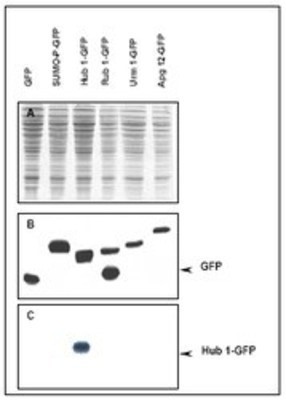
- Western Blot: UBL5 Antibody [NB600-470] - Immunoblot of Hub1 fusion protein. Anti-Hub1 antibody generated by immunization with recombinant yeast Hub1 was tested by immunoblot against yeast lysates expressing the Hub1-GFP fusion protein and other UBL fusion proteins. All UBLs possess limited homology to Ubiquitin and to each other, therefore it is important to know the degree of reactivity of each antibody against each UBL. Panel A shows total protein staining using ponceau. Panel B shows positions of free GFP or GFP containing recombinant proteins present in each lysate preparation after reaction with a 1:1,000 dilution of anti-GFP followed by reaction with a 1:15,000 dilution of HRP Donkey-a-Goat IgG MX. Panel C shows specific reaction with Hub1 using a 1:500 dilution of IgG fraction of Rabbit-anti-Hub1 (Yeast) followed by reaction with a 1:15,000 dilution of HRP Goat-a-Rabbit IgG MX. All primary antibodies were diluted in TTBS buffer supplemented with 5% non-fat milk and incubated with the membranes overnight at 4 degrees C. Yeast lysate proteins were separated by SDS-PAGE using a 15% gel. This data indicates that anti-Hub1 is highly specific and does not cross react with other UBLs. A chemiluminescence system was used for signal detection (Roche). Other detection systems will yield similar results. Data contributed by M. Malakhov, www.lifesensors.com, personal communication.
- Submitted by
- Novus Biologicals (provider)
- Main image
- Experimental details
- Western Blot: UBL5 Antibody [NB600-470] - Panel B shows positions of free GFP or GFP containing recombinant proteins present in each lysate preparation after reaction with a 1:1,000 dilution of anti-GFP followed by reaction with a 1:15,000 dilution of HRP Donkey-a-Goat IgG MX. Panel C shows specific reaction with Hub1 using a 1:500 dilution of IgG fraction of Rabbit-anti-Hub1 (Yeast) followed by reaction with a 1:15,000 dilution of HRP Goat-a-Rabbit IgG MX. All primary antibodies were diluted in TTBS buffer supplemented with 5% non-fat milk and incubated with the membranes overnight at 4 C. Yeast lysate proteins were separated by SDS-PAGE using a 15% gel. This data indicates that anti-Hub1 is highly specific and does not cross react with other UBLs.
