Explore
 Validate
 Learn
Learn Western blot
Antibody data
- Antibody Data
- Antigen structure
- References [3]
- Comments [0]
- Validations
- Western blot [1]
- Immunohistochemistry [1]
- Flow cytometry [1]
Submit
Validation data
Reference
Comment
Report error
- Product number
- AF2728 - Provider product page

- Provider
- Novus Biologicals
- Product name
- Goat Polyclonal SPARC-like 1/SPARCL1 Antibody
- Antibody type
- Polyclonal
Submitted references SPARCL1, a Novel Prognostic Predictive Factor for GI Malignancies: a Meta-Analysis.
Molecular resistance fingerprint of pemetrexed and platinum in a long-term survivor of mesothelioma.
Down-regulated SPARCL1 is associated with clinical significance in human gastric cancer.
Hu H, Cai W, Zheng S, Ge W
Cellular physiology and biochemistry : international journal of experimental cellular physiology, biochemistry, and pharmacology 2017;44(4):1485-1496
Cellular physiology and biochemistry : international journal of experimental cellular physiology, biochemistry, and pharmacology 2017;44(4):1485-1496
Molecular resistance fingerprint of pemetrexed and platinum in a long-term survivor of mesothelioma.
Røe OD, Szulkin A, Anderssen E, Flatberg A, Sandeck H, Amundsen T, Erlandsen SE, Dobra K, Sundstrøm SH
PloS one 2012;7(8):e40521
PloS one 2012;7(8):e40521
Down-regulated SPARCL1 is associated with clinical significance in human gastric cancer.
Li P, Qian J, Yu G, Chen Y, Liu K, Li J, Wang J
Journal of surgical oncology 2012 Jan;105(1):31-7
Journal of surgical oncology 2012 Jan;105(1):31-7
No comments: Submit comment
Supportive validation
- Submitted by
- Novus Biologicals (provider)
- Main image

- Experimental details
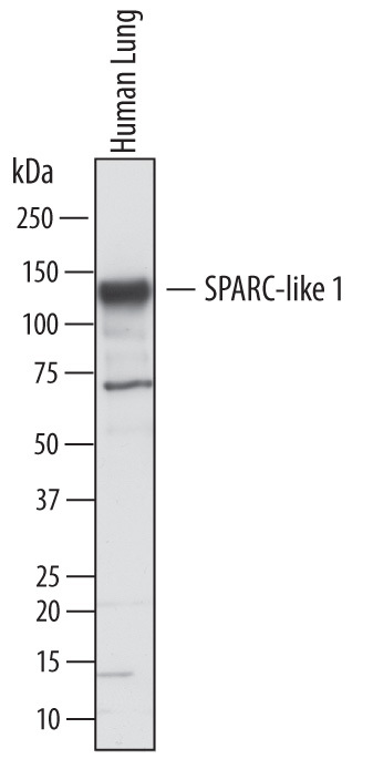
- Detection of Human SPARC-like 1/SPARCL1 by Western Blot. Western blot shows lysates of human lung tissue. PVDF membrane was probed with 1 µg/mL of Goat Anti-Human SPARC-like 1/ SPARCL1 Antigen Affinity-purified Polyclonal Antibody (Catalog # AF2728) followed by HRP-conjugated Anti-Goat IgG Secondary Antibody (Catalog # HAF017). A specific band was detected for SPARC-like 1/SPARCL1 at approximately 130 kDa (as indicated). This experiment was conducted under reducing conditions and using Immunoblot Buffer Group 1.
Supportive validation
- Submitted by
- Novus Biologicals (provider)
- Main image

- Experimental details
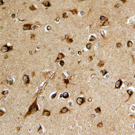
- SPARC-like 1/SPARCL1 in Human Brain. SPARC-like 1/SPARCL1 was detected in immersion fixed paraffin-embedded sections of human brain using Goat Anti-Human SPARC-like 1/SPARCL1 Antigen Affinity-purified Polyclonal Antibody (Catalog # AF2728) at 15 µg/mL overnight at 4 °C. Before incubation with the primary antibody, tissue was subjected to heat-induced epitope retrieval using Antigen Retrieval Reagent-Basic (Catalog # CTS013). Tissue was stained using the Anti-Goat HRP-DAB Cell & Tissue Staining Kit (brown; Catalog # CTS008) and counterstained with hematoxylin (blue). Specific staining was localized to neurons. View our protocol for Chromogenic IHC Staining of Paraffin-embedded Tissue Sections.
Supportive validation
- Submitted by
- Novus Biologicals (provider)
- Main image

- Experimental details
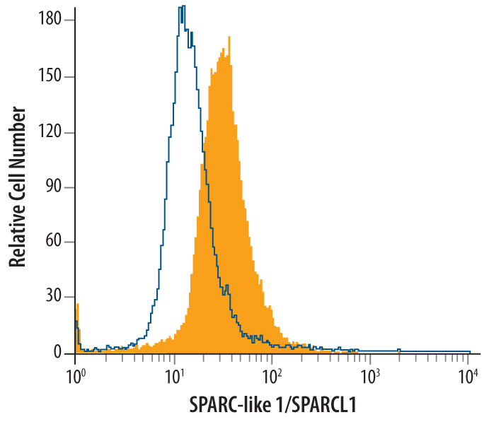
- Detection of SPARC-like 1/SPARCL1 in HL-60 Human Cell Line by Flow Cytometry. HL-60 human acute promyelocytic leukemia cell line was stained with Goat Anti-Human SPARC-like 1/SPARCL1 Antigen Affinity-purified Polyclonal Antibody (Catalog # AF2728, filled histogram) or control antibody (Catalog # AB-108-C, open histogram), followed by Phycoerythrin-conjugated Anti-Goat IgG Secondary Antibody (Catalog # F0107). To facilitate intracellular staining, cells were fixed with paraformaldehyde and permeabilized with saponin.