 Explore
 Validate
 Learn
Learn Western blot
Antibody data
- Antibody Data
- Antigen structure
- References [0]
- Comments [0]
- Validations
- Western blot [2]
- Immunocytochemistry [1]
- Immunohistochemistry [1]
Submit
Validation data
Reference
Comment
Report error
- Product number
- TA302073 - Provider product page

- Provider
- OriGene
- Product name
- Rabbit Polyclonal Antibody against PDHX (T11)
- Antibody type
- Polyclonal
- Description
- Rabbit Polyclonal Antibody against PDHX (T11)
- Host
- Rabbit
- Conjugate
- Unconjugated
- Epitope
- PDHX
- Antibody clone number
- NULL
- Vial size
- 100 µg
- Concentration
- 0.25 mg/ml
No comments: Submit comment
Supportive validation
- Submitted by
- OriGene (provider)
- Main image

- Experimental details
- Western blot analysis of anti-PDX1 Antibody (T11) (Cat.#TA302073) in NCI-H460 cell line lysates (35ug/lane).PDX1-T11(arrow) was detected using the purified Pab.
- Validation comment
- WB

- Submitted by
- OriGene (provider)
- Main image

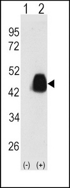
- Experimental details
- Western blot analysis of PDX1 (arrow) using PDX1 Antibody (T11) (Cat.#TA302073). 293 cell lysates (2 ug/lane) either nontransfected (Lane 1) or transiently transfected with the PDX1 gene (Lane 2) (Origene Technologies).
- Validation comment
- WB
Supportive validation
- Submitted by
- OriGene (provider)
- Main image

- Experimental details
- IF image of SY5Y cells stained with PDX1 (T11) antibody. SY5Y cells were incubated with TA302073 PDX1 (T11) primary antibody (1:100, 2 h at RT). For secondary antibody, Alexa Fluor? 488 conjugated donkey anti-rabbit antibody (green) was used (1:1000, 1h). Nuclei were counterstained with Hoechst 33342 (blue) . Note the highly specific localization of the PDX1 immunosignal to the nucleus, supported by Human Protein Atlas Data (http://www.proteinatlas.org/ENSG00000110435).
- Validation comment
- IF

Supportive validation
- Submitted by
- OriGene (provider)
- Main image

- Experimental details
- Formalin-fixed and paraffin-embedded human hepatocarcinoma tissue reacted with Phospho-PDX1-T11.ctrl antibody, which was peroxidase-conjugated to the secondary antibody, followed by DAB staining. This data demonstrates the use of this antibody for immunohistochemistry; clinical relevance has not been evaluated.
- Validation comment
- IHC
