 Explore
 Validate
 Learn
Learn Western blot
Antibody data
- Antibody Data
- Antigen structure
- References [1]
- Comments [0]
- Validations
- Western blot [1]
- Immunohistochemistry [2]
- Other assay [1]
Submit
Validation data
Reference
Comment
Report error
- Product number
- PA5-13725 - Provider product page

- Provider
- Invitrogen Antibodies
- Product name
- GRK1 Polyclonal Antibody
- Antibody type
- Polyclonal
- Antigen
- Synthetic peptide
- Reactivity
- Human, Mouse
- Host
- Rabbit
- Isotype
- IgG
- Vial size
- 400 µL
- Storage
- -20° C, Avoid Freeze/Thaw Cycles
Submitted references Sustained treatment of retinal vascular diseases with self-aggregating sunitinib microparticles.
Tsujinaka H, Fu J, Shen J, Yu Y, Hafiz Z, Kays J, McKenzie D, Cardona D, Culp D, Peterson W, Gilger BC, Crean CS, Zhang JZ, Kanan Y, Yu W, Cleland JL, Yang M, Hanes J, Campochiaro PA
Nature communications 2020 Feb 4;11(1):694
Nature communications 2020 Feb 4;11(1):694
No comments: Submit comment
Supportive validation
- Submitted by
- Invitrogen Antibodies (provider)
- Main image

- Experimental details
- Western blot analysis of GRK1 using a GRK1 polyclonal antibody (Product # PA5-13725) in HeLa cell lysate (Lane 1) and mouse spleen tissue lysate (Lane 2).

Supportive validation
- Submitted by
- Invitrogen Antibodies (provider)
- Main image

- Experimental details
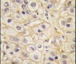
- Immunohistochemical analysis of formalin-fixed, paraffin-embedded human hepatocarcinoma tissue using a GRK1 polyclonal antibody (Product # PA5-13725), followed by HRP-conjugated secondary antibody and DAB staining.
- Submitted by
- Invitrogen Antibodies (provider)
- Main image

- Experimental details
- Immunohistochemical analysis of formalin-fixed, paraffin-embedded human cancer tissue using a GRK1 polyclonal antibody (Product # PA5-13725), followed by HRP-conjugated secondary antibody and DAB staining.

Supportive validation
- Submitted by
- Invitrogen Antibodies (provider)
- Main image
- Experimental details
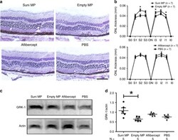
- Fig. 5 Intravitreous injection of sunitinib MPs in mice with type 3 CNV reduces photoreceptor cell death. Rho/VEGF mice were given an intravitreous injection of 10 ug of sunitinib microparticles (Suni MP) in one eye and 10 ug of empty MP in the other eye or 40 ug of aflibercept in one eye and PBS in the fellow eye. At P42, mice ( n = 7 for each group) were euthanized and serial frozen ocular sections were cut from the superior pole of the eye (S0) to the inferior pole (I0) and sections 25% (S1 and I1), 50% (S2 and I2), and 75% (S3 and I3) of the distance between each pole and the optic nerve (ON) were stained with hematoxylin and outer nuclear layer (ONL) thickness was measured by image analysis by a masked investigator. The ONL of sections from the S2 location of Suni MP-injected eyes appeared thicker than those from empty MP-injected eyes, but those from aflibercept-injected eyes appeared similar to those from PBS-injected eyes ( a scale bar = 100 um). The mean (+-SEM) ONL thickness was significantly greater at three of six locations in Suni MP-injected eyes compared with empty MP-injected eyes, but there was no difference between aflibercept- and PBS-injected eyes ( b ). At P49, mice ( n = 5 for each group) were euthanized and immunoblots of retinal homogenates from Suni MP-injected eyes showed prominent bands for rhodopsin kinase (GRK-1) ( c ). Denistometry showed that the mean (+-SEM) GRK-1/Actin ratio was significantly greater in retinas from Suni MP-injected eyes compa
