Explore
 Validate
 Learn
Learn Western blot
Antibody data
- Antibody Data
- Antigen structure
- References [0]
- Comments [0]
- Validations
- Western blot [1]
- Immunocytochemistry [1]
- Immunohistochemistry [1]
- Flow cytometry [1]
Submit
Validation data
Reference
Comment
Report error
- Product number
- NBP2-29948 - Provider product page

- Provider
- Novus Biologicals
- Product name
- Rabbit Polyclonal Ext2 Antibody
- Antibody type
- Polyclonal
- Description
- Protein A purified.
- Reactivity
- Human
- Host
- Rabbit
- Isotype
- IgG
- Vial size
- 0.4 ml
- Concentration
- 0.3 mg/ml
- Storage
- Store at 4C short term. Aliquot and store at -20C long term. Avoid freeze-thaw cycles.
No comments: Submit comment
Supportive validation
- Submitted by
- Novus Biologicals (provider)
- Main image
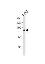
- Experimental details
- Western Blot: Ext2 Antibody [NBP2-29948] - Western blot analysis of lysate from T47D cell line,using EXT2 Antibody (Center)(NBP2-29948).AP9401c was diluted at 1:1000 at each lane. A goat anti-rabbit IgG H&L(HRP) at 1:5000 dilution was used as the secondary antibody.Lysate at 35ug per lane.

Supportive validation
- Submitted by
- Novus Biologicals (provider)
- Main image

- Experimental details
- Immunocytochemistry/Immunofluorescence: Ext2 Antibody [NBP2-29948] - Confocal immunofluorescent analysis of EXT2 Antibody (Center)(NBP2-29948) with hela cell followed by Alexa Fluor 488-conjugated goat anti-rabbit lgG (green). DAPI was used to stain the cell nuclear (blue).

Supportive validation
- Submitted by
- Novus Biologicals (provider)
- Main image

- Experimental details
- Immunohistochemistry-Paraffin: Ext2 Antibody [NBP2-29948] - Formalin-fixed and paraffin-embedded human brain tissue reacted with EXT2 Antibody (Center), which was peroxidase-conjugated to the secondary antibody, followed by DAB staining. This data demonstrates the use of this antibody for immunohistochemistry; clinical relevance has not been evaluated.

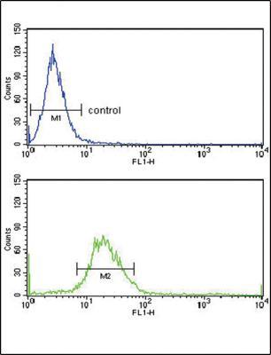
Supportive validation
- Submitted by
- Novus Biologicals (provider)
- Main image

- Experimental details
- Flow Cytometry: Ext2 Antibody [NBP2-29948] - Flow cytometry analysis of Hela cells (bottom histogram) compared to a negative control cell (top histogram).FITC-conjugated goat-anti-rabbit secondary antibodies were used for the analysis.