Explore
 Validate
 Learn
Learn Western blot
Antibody data
- Antibody Data
- Antigen structure
- References [0]
- Comments [0]
- Validations
- Western blot [1]
- Immunocytochemistry [1]
- Immunohistochemistry [2]
Submit
Validation data
Reference
Comment
Report error
- Product number
- NBP1-03361 - Provider product page

- Provider
- Novus Biologicals
- Proper citation
- Novus Cat#NBP1-03361, RRID:AB_1522375
- Product name
- Rabbit Polyclonal Shugoshin Antibody
- Antibody type
- Polyclonal
- Description
- Immunogen affinity purified.
- Reactivity
- Human
- Host
- Rabbit
- Isotype
- IgG
- Vial size
- 0.1 ml
- Concentration
- 1 mg/ml
- Storage
- Store at 4C. Do not freeze.
No comments: Submit comment
Supportive validation
- Submitted by
- Novus Biologicals (provider)
- Main image

- Experimental details
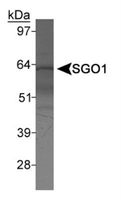
- Western Blot: Shugoshin Antibody [NBP1-03361] - Detection of SGO1 in HeLa nuclear extract using NBP1-03361.
Supportive validation
- Submitted by
- Novus Biologicals (provider)
- Main image

- Experimental details
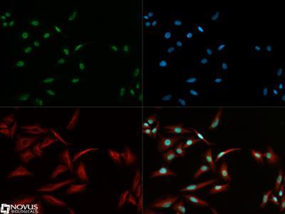
- Immunocytochemistry/Immunofluorescence: Shugoshin Antibody [NBP1-03361] - Shugoshin antibody was tested in HeLa cells with DyLight 488 (green). Nuclei and alpha-tubulin were counterstained with DAPI (blue) and Dylight 550 (red).
Supportive validation
- Submitted by
- Novus Biologicals (provider)
- Main image
- Experimental details
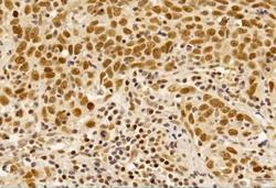
- Immunohistochemistry-Paraffin: Shugoshin Antibody [NBP1-03361] - IHC analysis of formalin-fixed paraffin-embedded tissue section of human pulmonary squamous cell carcinoma using rabbit polyclonal Shugoshin antibody (NBP1-03361) at 5 ug/ml concentration. The carcinoma cells depicted a distinct nuclear with weak cytoplasmic immuno-reactivity for Shugoshin protein [Magnification 40X]
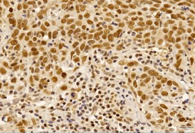
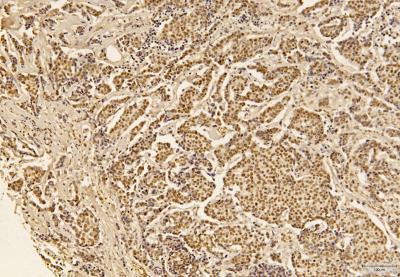
- Submitted by
- Novus Biologicals (provider)
- Main image

- Experimental details
- Immunohistochemistry-Paraffin: Shugoshin Antibody [NBP1-03361] - IHC analysis of formalin-fixed paraffin-embedded tissue section of infiltrating ductal carcinoma of human breast using rabbit polyclonal Shugoshin antibody (NBP1-03361) at 5 ug/ml concentration. The carcinoma cells depicted a moderate to strong nuclear with weak cytoplasmic immunopositivity of Shugoshin protein, whereas, the collagen fibers in the surrounding tumor stroma/connective tissue also developed a very weak/potentially nonspecific staining [Magnification 40X].