Explore
 Validate
 Learn
Learn Western blot
 Immunocytochemistry
Antibody data
- Antibody Data
- Antigen structure
- References [4]
- Comments [0]
- Validations
- Western blot [3]
- Immunoprecipitation [1]
- Immunohistochemistry [6]
- Flow cytometry [2]
Submit
Validation data
Reference
Comment
Report error
- Product number
- NBP2-16908 - Provider product page

- Provider
- Novus Biologicals
- Product name
- Rabbit Polyclonal AIF-1/Iba1 Antibody
- Antibody type
- Polyclonal
- Description
- Immunogen affinity purified.
- Reactivity
- Human, Mouse, Rat
- Host
- Rabbit
- Isotype
- IgG
- Vial size
- 0.1 ml
- Storage
- Aliquot and store at -20C or -80C. Avoid freeze-thaw cycles.
Submitted references Devising micro/nano-architectures in multi-channel nerve conduits towards a pro-regenerative matrix for the repair of spinal cord injury.
Annexin A1-derived peptide Ac(2-26) in a pilocarpine-induced status epilepticus model: anti-inflammatory and neuroprotective effects.
Pharmacogenetic neuronal stimulation increases human tau pathology and trans-synaptic spread of tau to distal brain regions in mice.
Purinergic P2Y1 Receptors Control Rapid Expression of Plasma Membrane Processes in Hippocampal Astrocytes.
Sun X, Bai Y, Zhai H, Liu S, Zhang C, Xu Y, Zou J, Wang T, Chen S, Zhu Q, Liu X, Mao H, Quan D
Acta biomaterialia 2019 Mar 1;86:194-206
Acta biomaterialia 2019 Mar 1;86:194-206
Annexin A1-derived peptide Ac(2-26) in a pilocarpine-induced status epilepticus model: anti-inflammatory and neuroprotective effects.
Gimenes AD, Andrade BFD, Pinotti JVP, Oliani SM, Galvis-Alonso OY, Gil CD
Journal of neuroinflammation 2019 Feb 12;16(1):32
Journal of neuroinflammation 2019 Feb 12;16(1):32
Pharmacogenetic neuronal stimulation increases human tau pathology and trans-synaptic spread of tau to distal brain regions in mice.
Schultz MK Jr, Gentzel R, Usenovic M, Gretzula C, Ware C, Parmentier-Batteur S, Schachter JB, Zariwala HA
Neurobiology of disease 2018 Oct;118:161-176
Neurobiology of disease 2018 Oct;118:161-176
Purinergic P2Y1 Receptors Control Rapid Expression of Plasma Membrane Processes in Hippocampal Astrocytes.
Chisari M, Scuderi A, Ciranna L, Volsi GL, Licata F, Sortino MA
Molecular neurobiology 2017 Aug;54(6):4081-4093
Molecular neurobiology 2017 Aug;54(6):4081-4093
No comments: Submit comment
Supportive validation
- Submitted by
- Novus Biologicals (provider)
- Main image

- Experimental details
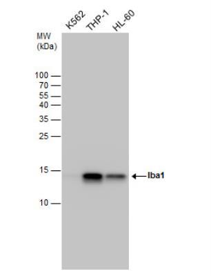
- Western Blot: AIF-1/Iba1 Antibody [NBP2-16908] - Various whole 30 ug cell extracts (Molecular weight: 16.7 KDa) were separated by 15% SDS-PAGE, and the membrane was blotted with AIF-1/Iba1 Antibody diluted at a dilution of 1:5000.
- Submitted by
- Novus Biologicals (provider)
- Main image

- Experimental details
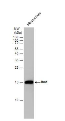
- Western Blot: AIF-1/Iba1 Antibody [NBP2-16908] - Mouse tissue extracts (50 ug) was separated by 15 % SDS-PAGE, and the membrane was blotted with AIF-1/Iba1 Antibody diluted by 1:500.
- Submitted by
- Novus Biologicals (provider)
- Main image

- Experimental details
- Western Blot: AIF-1/Iba1 Antibody [NBP2-16908] - Rat tissue extract (50 ug) was separated by 15% SDS-PAGE, and the membrane was blotted with AIF-1/Iba1 Antibody diluted at 1:1000. The HRP-conjugated anti-rabbit IgG antibody (NBP2-19301) was used to detect the primary antibody.

Supportive validation
- Submitted by
- Novus Biologicals (provider)
- Main image
- Experimental details
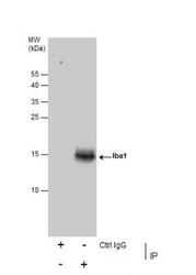
- Immunoprecipitation: AIF-1/Iba1 Antibody [NBP2-16908] - Immunoprecipitation of Iba1 protein from K562 whole cell extracts using 5ug of Iba1 antibody Western blot analysis was performed using Iba1 antibody EasyBlot anti-Rabbit IgG was used as a secondary reagent.

Supportive validation
- Submitted by
- Novus Biologicals (provider)
- Main image

- Experimental details
- Immunohistochemistry-Paraffin: AIF-1/Iba1 Antibody [NBP2-16908] - Paraffin-embedded breast cancer stroma. Iba1 antibody dilution: 1:500.

- Submitted by
- Novus Biologicals (provider)
- Main image

- Experimental details
- Immunohistochemistry-Frozen: AIF-1/Iba1 Antibody [NBP2-16908] - Frozen sectioned E13.5 Rat brain. Green: AIF-1/Iba1 protein stained by AIF-1/Iba1 Antibody diluted at 1:250. Red: beta Tubulin 3/ TUJ1, a mature neuron marker, stained by beta Tubulin 3/ TUJ1 antibody [11710] (NBP2-43559) diluted at 1:500. Blue: Fluoroshield with DAPI.

- Submitted by
- Novus Biologicals (provider)
- Main image

- Experimental details
- Immunohistochemistry-Paraffin: AIF-1/Iba1 Antibody [NBP2-16908] - Paraffin-embedded mouse brain.Iba1 antibody diluted at 1:500.

- Submitted by
- Novus Biologicals (provider)
- Main image

- Experimental details
- Immunohistochemistry-Paraffin: AIF-1/Iba1 Antibody [NBP2-16908] - Paraffin-embedded rat brain stained using AIF-1/Iba1 Antibody diluted at 1:500.

- Submitted by
- Novus Biologicals (provider)
- Main image

- Experimental details
- Immunohistochemistry-Paraffin: AIF-1/Iba1 Antibody [NBP2-16908] - Mouse thymus gland stained by AIF-1/Iba1 Antibody diluted at 1:500. Antigen Retrieval: Citrate buffer, pH 6.0, 15 min

- Submitted by
- Novus Biologicals (provider)
- Main image

- Experimental details
- Immunohistochemistry-Paraffin: AIF-1/Iba1 Antibody [NBP2-16908] - Mouse brain stained by AIF-1/Iba1 Antibody diluted at 1:500. Antigen Retrieval: Citrate buffer, pH 6.0, 15 min

Supportive validation
- Submitted by
- Novus Biologicals (provider)
- Main image

- Experimental details
- Flow Cytometry: AIF-1/Iba1 Antibody [NBP2-16908] - Primary murine microglia cells, staining with AIF-1:Iba1 Antibody using 1. 0 ug per 4 x 10^5 cells. NBP2-16908 (blue), Rabbit IgG (green) Unstained (red).

- Submitted by
- Novus Biologicals (provider)
- Main image

- Experimental details
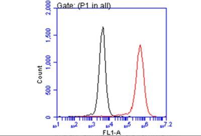
- Flow Cytometry: AIF-1/Iba1 Antibody [NBP2-16908] - THP-1 cell. Black: Unlabelled sample was used as a control. Red: AIF-1:Iba1 Antibody dilution: 1:50. Acquisition of 20,000 events were collected using a Dylight 488-conjugated secondary antibody for FACS analysis.