 Explore
 Validate
 Learn
Learn Western blot
 Immunocytochemistry
Antibody data
- Antibody Data
- Antigen structure
- References [6]
- Comments [0]
- Validations
- Western blot [8]
- Immunoprecipitation [1]
- Immunohistochemistry [4]
- Flow cytometry [2]
Submit
Validation data
Reference
Comment
Report error
- Product number
- GTX101495 - Provider product page

- Provider
- GeneTex
- Proper citation
- GeneTex Cat#GTX101495, RRID:AB_1240433
- Product name
- Iba1 antibody
- Antibody type
- Polyclonal
- Reactivity
- Human, Mouse, Rat
- Host
- Rabbit
Submitted references Surfactant protein A is expressed in the central nervous system of rats with experimental autoimmune encephalomyelitis, and suppresses inflammation in human astrocytes and microglia.
A vaccine with Aβ oligomer-specific mimotope attenuates cognitive deficits and brain pathologies in transgenic mice with Alzheimer's disease.
A new animal model containing human SCARB2 and lacking stat-1 is highly susceptible to EV71.
A Novel Multifunctional Compound Camellikaempferoside B Decreases Aβ Production, Interferes with Aβ Aggregation, and Prohibits Aβ-Mediated Neurotoxicity and Neuroinflammation.
Targeting P(2)X(7) receptor for the treatment of central post-stroke pain in a rodent model.
Immunodeficient mouse models with different disease profiles by in vivo infection with the same clinical isolate of enterovirus 71.
Yang X, Yan J, Feng J
Molecular medicine reports 2017 Jun;15(6):3555-3565
Molecular medicine reports 2017 Jun;15(6):3555-3565
A vaccine with Aβ oligomer-specific mimotope attenuates cognitive deficits and brain pathologies in transgenic mice with Alzheimer's disease.
Wang SW, Liu DQ, Zhang LX, Ji M, Zhang YX, Dong QX, Liu SY, Xie XX, Liu RT
Alzheimer's research & therapy 2017 Jun 7;9(1):41
Alzheimer's research & therapy 2017 Jun 7;9(1):41
A new animal model containing human SCARB2 and lacking stat-1 is highly susceptible to EV71.
Liou AT, Wu SY, Liao CC, Chang YS, Chang CS, Shih C
Scientific reports 2016 Aug 8;6:31151
Scientific reports 2016 Aug 8;6:31151
A Novel Multifunctional Compound Camellikaempferoside B Decreases Aβ Production, Interferes with Aβ Aggregation, and Prohibits Aβ-Mediated Neurotoxicity and Neuroinflammation.
Yang S, Liu W, Lu S, Tian YZ, Wang WY, Ling TJ, Liu RT
ACS chemical neuroscience 2016 Apr 20;7(4):505-18
ACS chemical neuroscience 2016 Apr 20;7(4):505-18
Targeting P(2)X(7) receptor for the treatment of central post-stroke pain in a rodent model.
Kuan YH, Shih HC, Tang SC, Jeng JS, Shyu BC
Neurobiology of disease 2015 Jun;78:134-45
Neurobiology of disease 2015 Jun;78:134-45
Immunodeficient mouse models with different disease profiles by in vivo infection with the same clinical isolate of enterovirus 71.
Liao CC, Liou AT, Chang YS, Wu SY, Chang CS, Lee CK, Kung JT, Tu PH, Yu YY, Lin CY, Lin JS, Shih C
Journal of virology 2014 Nov;88(21):12485-99
Journal of virology 2014 Nov;88(21):12485-99
No comments: Submit comment
Supportive validation
- Submitted by
- GeneTex (provider)
- Main image

- Experimental details
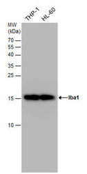
- Sample (30 ug of whole cell lysate) A: THP-1 B: HL-60 15% SDS PAGE GTX101495 diluted at 1:5000
- Validation comment
- WB

- Submitted by
- GeneTex (provider)
- Main image

- Experimental details
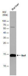
- Sample (50 ug of whole cell lysate) A: mouse Liver 15% SDS PAGE GTX101495 diluted at 1:1000
- Validation comment
- WB

- Submitted by
- GeneTex (provider)
- Main image

- Experimental details
- Iba1 antibody detects Iba1 protein by Western blot analysis. Mouse tissue extracts (50 µg) was separated by 15 % SDS-PAGE, and the membrane was blotted with Iba1 antibody (GTX101495) diluted by 1:500.
- Validation comment
- WB

- Submitted by
- GeneTex (provider)
- Main image

- Experimental details
- Iba1 antibody detects Iba1 protein by western blot analysis. Various whole cell extracts (30 ?g) were separated by 15% SDS-PAGE, and blotted with Iba1 antibody (GTX101495) diluted by 1:2500. The HRP-conjugated anti-rabbit IgG antibody (GTX213110-01) was used to detect the primary antibody.

- Submitted by
- GeneTex (provider)
- Main image
- Experimental details
- Iba1 antibody detects Iba1 protein by western blot analysis. Various whole cell extracts (30 ?g) were separated by 15 % SDS-PAGE, and blotted with Iba1 antibody (GTX101495) diluted by 1:2500
- Validation comment
- WB

- Submitted by
- GeneTex (provider)
- Main image

- Experimental details
- Iba1 antibody detects Iba1 protein by western blot analysis. Various whole cell extracts (30 ?g) were separated by 15% SDS-PAGE, and the membrane was blotted with Iba1 antibody (GTX101495) diluted at a dilution of 1:5000. The HRP-conjugated anti-rabbit IgG antibody (GTX213110-01) was used to detect the primary antibody.

- Submitted by
- GeneTex (provider)
- Main image

- Experimental details
- Mouse tissue extract (50 ?g) was separated by 15% SDS-PAGE, and the membrane was blotted with Iba1 antibody (GTX101495) diluted at 1:1000. The HRP-conjugated anti-rabbit IgG antibody (GTX213110-01) was used to detect the primary antibody.

- Submitted by
- GeneTex (provider)
- Main image
- Experimental details
- Rat tissue extract (50 ?g) was separated by 15% SDS-PAGE, and the membrane was blotted with Iba1 antibody (GTX101495) diluted at 1:1000. The HRP-conjugated anti-rabbit IgG antibody (GTX213110-01) was used to detect the primary antibody.

Supportive validation
- Submitted by
- GeneTex (provider)
- Main image

- Experimental details
- Immunoprecipitation of Iba1 protein from K562 whole cell extracts using 5 £gg of Iba1 antibody (GTX101495).Western blot analysis was performed using Iba1 antibody (GTX101495).EasyBlot anti-Rabbit IgG (GTX221666-01) was used as a secondary reagent.

Supportive validation
- Submitted by
- GeneTex (provider)
- Main image
- Experimental details
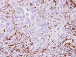
- Iba1 antibody detects Iba1 protein at cytoplasm on human breast cancer stroma by immunohistochemical analysis. Sample: Paraffin-embedded breast cancer stroma. Iba1 antibody (GTX101495) dilution: 1:500.

- Submitted by
- GeneTex (provider)
- Main image

- Experimental details
- Iba1 antibody detects Iba1 protein expression at microglias by immunohistochemical analysis.Sample: Frozen sectioned E13.5 Rat brain. Green: Iba1 protein stained by Iba1 antibody (GTX101495) diluted at 1:250.Red: beta Tubulin 3/ TUJ1, a mature neuron marker, stained by beta Tubulin 3/ TUJ1 antibody [GT11710] (GTX631836) diluted at 1:500.Blue: Fluoroshield with DAPI (GTX30920).

- Submitted by
- GeneTex (provider)
- Main image

- Experimental details
- Iba1 antibody detects Iba1 protein at microglia in mouse brain by immunohistochemical analysis. Sample: Paraffin-embedded mouse brain. Iba1 antibody (GTX101495) diluted at 1:500.

- Submitted by
- GeneTex (provider)
- Main image

- Experimental details
- Iba1 antibody detects Iba1 protein at microglia in rat brain by immunohistochemical analysis. Sample: Paraffin-embedded rat brain. Iba1 antibody (GTX101495) diluted at 1:500.

Supportive validation
- Submitted by
- GeneTex (provider)
- Main image
- Experimental details
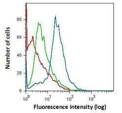
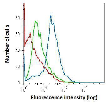
- Flow cytometry on primary murine microglia cells, staining with Iba1 (GTX101495) antibody using 1.0 ?g per 4¡Ñ105 cells. GTX101495 (blue), Rabbit IgG (green) ,Unstained (red).
- Submitted by
- GeneTex (provider)
- Main image

- Experimental details
- Iba1 antibody (GTX101495) detects Iba1 by flow cytometry analysis. Sample: THP-1 cell. Black: Unlabelled sample was used as a control. Red: Iba1 antibody (GTX101495) dilution: 1:50.
