Explore
 Validate
 Learn
Learn Immunocytochemistry
 Immunohistochemistry
Antibody data
- Antibody Data
- Antigen structure
- References [0]
- Comments [0]
- Validations
- Immunocytochemistry [1]
- Immunohistochemistry [19]
Submit
Validation data
Reference
Comment
Report error
- Product number
- HPA049117 - Provider product page

- Provider
- Atlas Antibodies
- Proper citation
- Atlas Antibodies Cat#HPA049117, RRID:AB_2680639
- Product name
- Anti-BCL11B
- Antibody type
- Polyclonal
- Reactivity
- Human, Mouse
- Host
- Rabbit
- Conjugate
- Unconjugated
- Antigen sequence
QGNPQHLSQRELITPEADHVEAAILEEDEGLEIEE
PSGLGLMVGGPDPDLLTCG- Isotype
- IgG
- Vial size
- 100 µl
- Storage
- Store at +4°C for short term storage. Long time storage is recommended at -20°C.
No comments: Submit comment
Supportive validation
- Submitted by
- Atlas Antibodies (provider)
- Main image
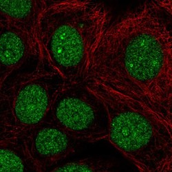
- Experimental details
- Immunofluorescent staining of human cell line MCF7 shows localization to nucleus & nucleoli fibrillar center.
- Sample type
- HUMAN

Enhanced validation
Supportive validation
- Submitted by
- Atlas Antibodies (provider)
- Enhanced method
- Orthogonal validation
- Main image

- Experimental details
- Immunohistochemistry analysis in human skin and pancreas tissues using HPA049117 antibody. Corresponding BCL11B RNA-seq data are presented for the same tissues.
- Sample type
- HUMAN

Supportive validation
- Submitted by
- Atlas Antibodies (provider)
- Main image
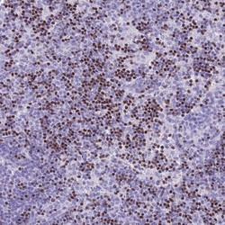
- Experimental details
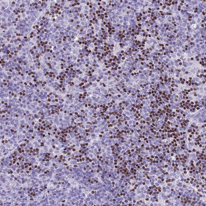
- Immunohistochemical staining of human lymph node shows strong nuclear positivity in subsets of non-germinal center cells.
- Submitted by
- Atlas Antibodies (provider)
- Main image
- Experimental details
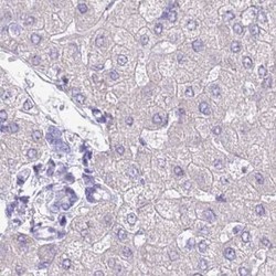
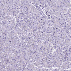
- liver
- Submitted by
- Atlas Antibodies (provider)
- Main image

- Experimental details
- liver cancer
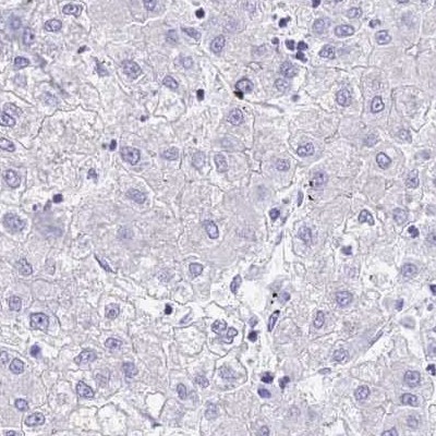
- Submitted by
- Atlas Antibodies (provider)
- Main image

- Experimental details
- Immunofluorescence staining of mouse basal forebrain shows nuclear positivity in neurons of caudate putamen.
- Submitted by
- Atlas Antibodies (provider)
- Main image
- Experimental details
- Immunohistochemical staining of human lateral ventricle shows nuclear positivity in subsets of neurons.

- Submitted by
- Atlas Antibodies (provider)
- Main image

- Experimental details
- Immunofluorescence staining of mouse olfactory bulb shows nuclear immunoreactivity in a subset of neurons.

- Submitted by
- Atlas Antibodies (provider)
- Main image

- Experimental details
- Immunofluorescence staining of mouse nucleus accumbens shows nuclear positivity in neurons.
- Submitted by
- Atlas Antibodies (provider)
- Main image
- Experimental details
- Immunofluorescence staining of mouse parietal association cortex shows nuclear immunoreactivity in neurons.

- Submitted by
- Atlas Antibodies (provider)
- Main image
- Experimental details
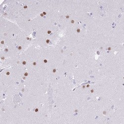
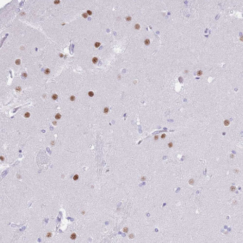
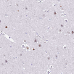
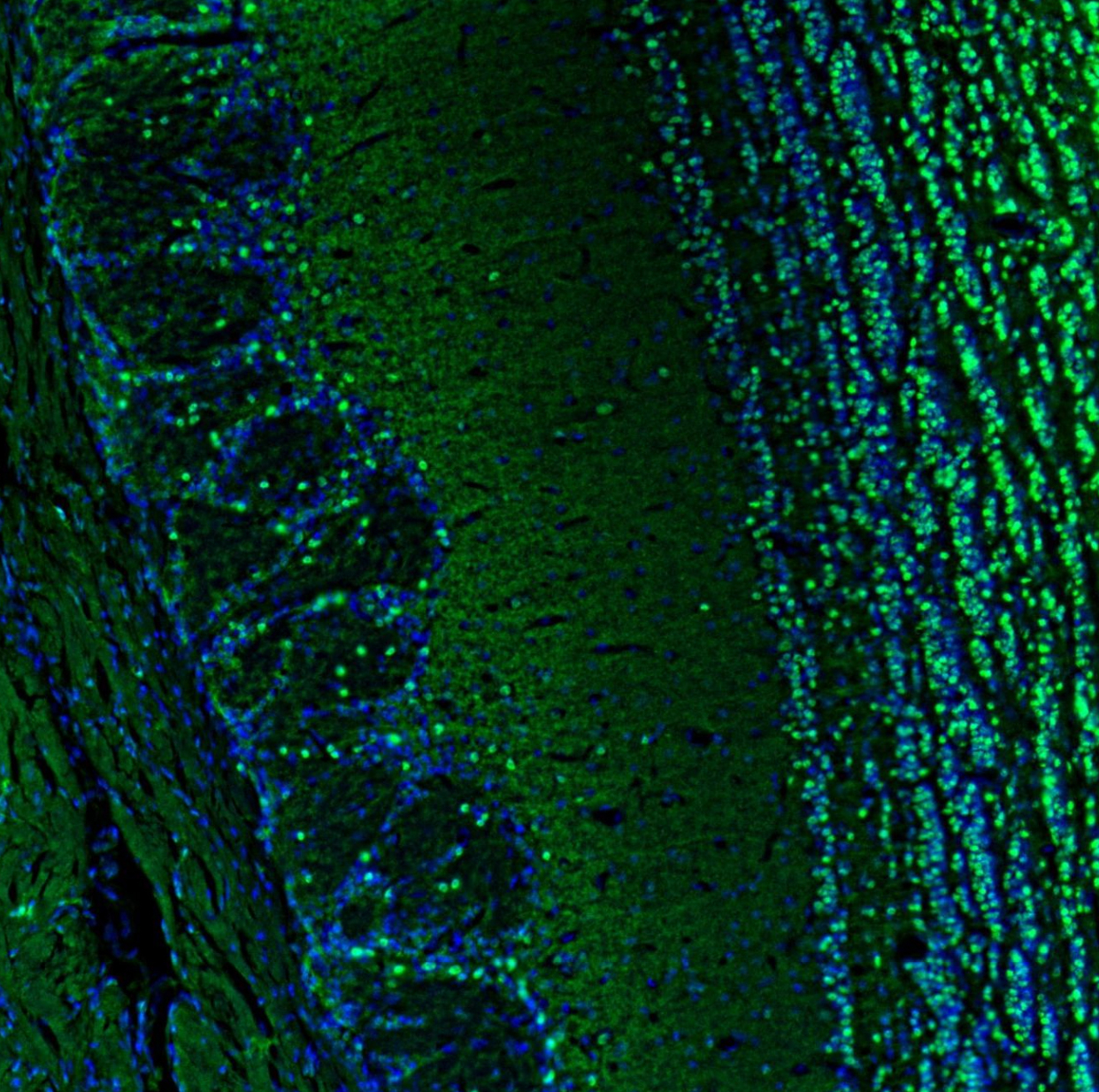
- Immunohistochemical staining of human cerebral cortex shows strong nuclear positivity in a subset of neurons.

- Submitted by
- Atlas Antibodies (provider)
- Main image
- Experimental details
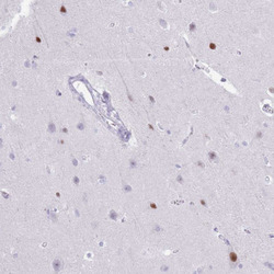
- Immunohistochemical staining of human lateral ventricle wall shows nuclear immunoreactivity in neurons.
- Submitted by
- Atlas Antibodies (provider)
- Main image
- Experimental details
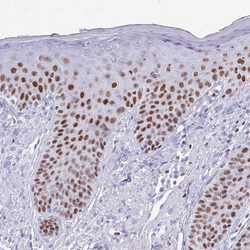
- Immunohistochemical staining of human skin shows moderate to strong nuclear positivity in epidermal cells.
- Sample type
- HUMAN

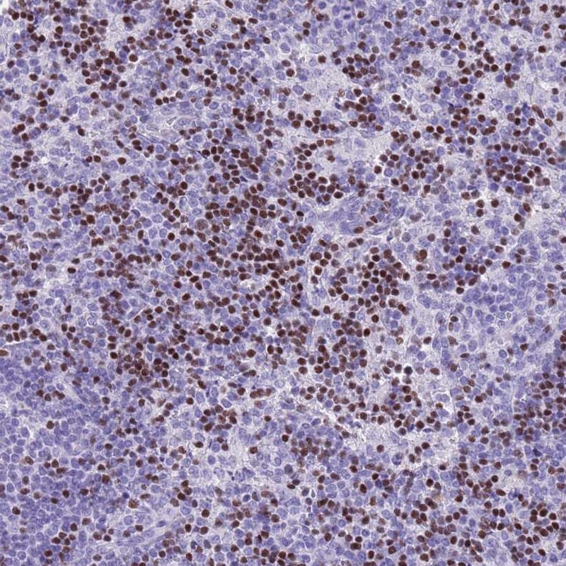
- Submitted by
- Atlas Antibodies (provider)
- Main image

- Experimental details
- Immunohistochemical staining of human lymph node shows moderate to strong nuclear positivity in non - germinal center cells.
- Sample type
- HUMAN
- Submitted by
- Atlas Antibodies (provider)
- Main image
- Experimental details
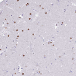
- Immunohistochemical staining of human cerebral cortex shows weak to moderate nuclear positivity in neurons.
- Sample type
- HUMAN

- Submitted by
- Atlas Antibodies (provider)
- Main image
- Experimental details
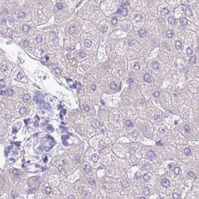
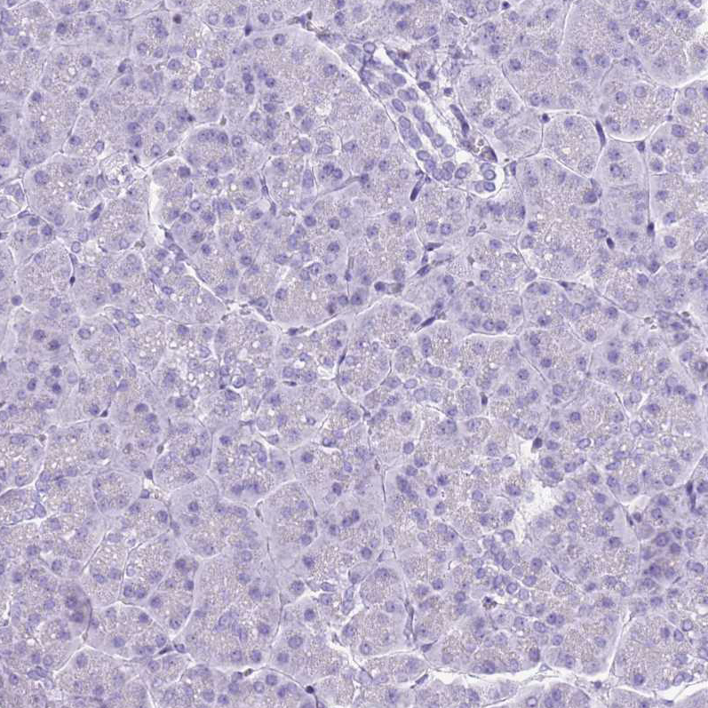
- Immunohistochemical staining of human pancreas shows no nuclear positivity in exocrine glandular cells as expected.
- Sample type
- HUMAN
- Submitted by
- Atlas Antibodies (provider)
- Main image
- Experimental details
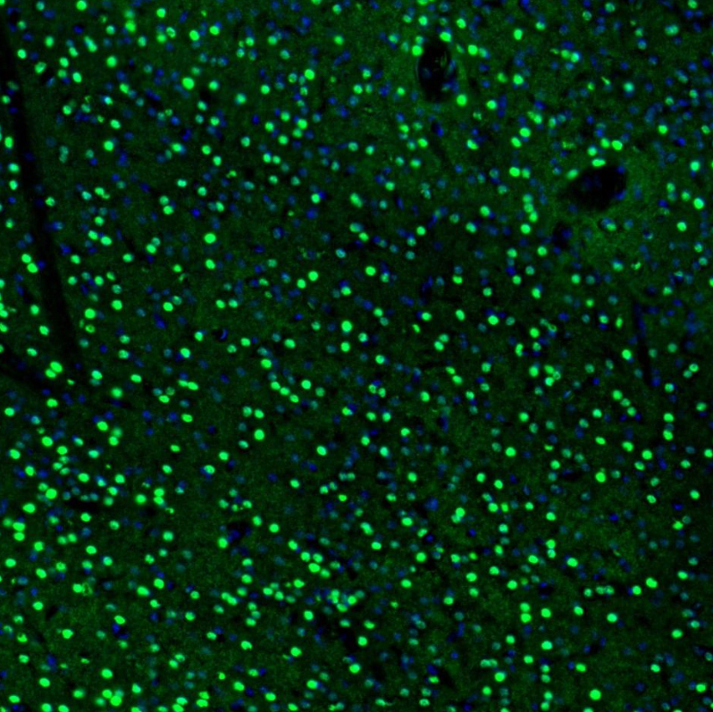
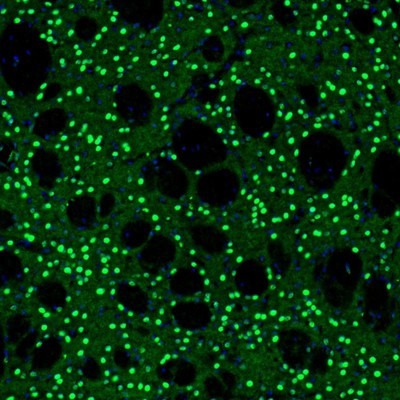
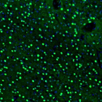
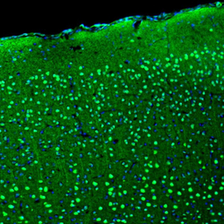
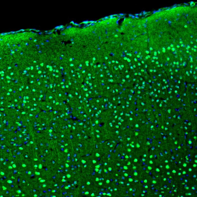
- Immunofluorescence staining of mouse brain shows moderate to strong positivity in neuronal cells in the cerebral cortex.
- Sample type
- MOUSE
- Submitted by
- Atlas Antibodies (provider)
- Main image

- Experimental details
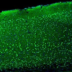
- Immunofluorescence staining of mouse brain shows moderate to strong positivity in neurons in the olfactory bulb.
- Sample type
- MOUSE
- Submitted by
- Atlas Antibodies (provider)
- Main image

- Experimental details
- Immunofluorescence staining of mouse basal forebrain shows moderate to strong positivity in neurons in the caudate putamen.
- Sample type
- MOUSE

- Submitted by
- Atlas Antibodies (provider)
- Main image

- Experimental details
- Immunofluorescence staining of mouse basal forebrain shows strong positivity in neurons in the accumbens nucleus, shell.
- Sample type
- MOUSE